

Authors
|
|||||||
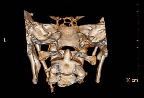
AbstractEagle Syndrome is characterized by ear, throat and neck pain, swallowing difficulty, and foreign body sensation in the throat due to styloid process elongation or stylohyoid ligament calcification. The diagnosis is made by palpation of styloid process at the tonsillar fossa and by three-dimensional computed tomography. The treatment is usually surgical excision. In this case report, a 51-year-old male with the diagnosis of Eagle's syndrome, who presented with complaints of otalgia and throat pain radiating to the neck during swallowing is presented and discussed in regard of current literature.IntroductionEagle Syndrome is characterized by stylohyoid ligament calcification or styloid process elongation causing irritation-related symptoms during chewing and swallowing [1,2]. Irritation-related symptoms are a painful sensation as a result of the pressure effect on various structures in the head and neck region. Symptoms commonly include odynophagia, dysphagia, tinnitus, cervicofacial pain, swallowing difficulty and globus or a foreign body sensation [3]. It is usually seen in women over 50 years old. Although the pathology is often seen bilaterally, symptoms are usually unilateral. The elongated styloid process and clinical findings were first defined by Eagle in 1937 [4] and estimated frequency of this syndrome is 4-8 per 10,000 people in the community [5]. However, only 4% of these are symptomatic [1]. The etiopathogenesis of Eagle syndrome is still unclear. It can be idiopathic, acquired due to trauma such as tonsillectomy or chronic irritation of the stylomandibular ligament may cause reactive ossificant hyperplasia of the styloid process or congenital due to the presence of persistence mesenchymal elements [Reichert cartilage residues] producing bone tissue of styloid ligament [4,6,7]. Diagnosis of Eagle Syndrome is made both by physical examination and by imaging methods. Palpation of the styloid process at the tonsil bed and pain during palpation is highly suspicious for Eagle syndrome. Imaging methods such as anterior-posterior direct x-ray, panoramic x-ray, computerized tomography [CT] can be used in diagnosis of the patients with Eagle syndrome. Three-dimensional CT [3D-CT] is an extremely valuable imaging tool for offering an accurate evaluation of the styloid process in relation with regional head and neck anatomy and also for surgical planning [8]. In treatment of Eagle syndrome, analgesics can be offered for pain alleviation. However, usually surgical excision either via intraoral approach or cervical approach may be performed for further treatment [9]. The target of this study is to discuss clinical presentation and also surgical therapy via a transoral approach of a case of patient diagnosed of Eagle syndrome. Case ReportA 51-year-old male patient who complained of left otalgia and throat pain radiating to the neck during swallowing for one year was admitted to our department. He had been seen in different clinics many times and received many medical treatment with no use. Since any pathology causing otalgia was not detected in the detailed otolaryngology examination, Eagle syndrome was suspected. A 3D-CT scan of the styloid process was performed for diagnosis. The right styloid process length was reported to be 24 mm within normal limits, however the left styloid process was reported as approximately 65 mm evaluated as significantly elongated with extension to left parapharyngeal fatty planes at the level of oropharynx (Figure 1-2). The findings were consistent with Eagle's syndrome. The patient was diagnosed with left-sided Eagle syndrome and surgery via transoral approach was recommended.
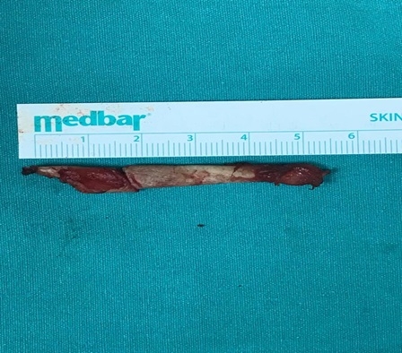
The left elongated styloid process was excised through transoral approach without tonsillectomy (Figure 3). During surgery, following orotracheal intubation under general anesthesia, the most distant part of styloid process was palpated at tonsil region and an incision of about 3 cm was done 1 cm lateral to the free margin of the anterior tonsilar pillar. The internal carotid artery was palpated and the muscles and ligaments adhering to the bone were separated by the elevator while paying attention to the arteries. The extreme part was held with an angled clamp and the distal part approximately 6 cm from distal styloid processes was excised respectively. After excision of subtotal part of elongated styloid process, the operation site was washed with saline, the insicion was closed with 3/0 vicryl. There were no peroperative or postoperative complications. In the second postoperative week the complaints of the patient was resolved.
DiscussionThe styloid process is a cylindrical shaped bone extension located in front of the stylomastoid foramen at the inferior aspect of the temporal bone. All stylohyoid and stylomandibular ligaments and also stylopharyngeus, stylohyoid and styloglossus muscles attach to this process. Stylohyoid ligament is the continuation of the styloid process and inserts to the lesser horn of the hyoid bone [10,11]. In cadaver studies, the normal length of the styloid process was found to be 20-30 mm varying on average [12]. Usually length of styloid processes and/or stylohyoid ligaments longer than 30 mm are accepted as an elongated styloid process [13,14,15,16]. In our case the right styloid process was 24 mm, while the left styloid process was elongated and was approximately 65 mm, significantly longer than normal. The frequency of Eagle Syndrome is 4% in population, but only 4 to 10.3% of these patients are symptomatic [1,8]. There is a female predominance and it is common over 50 years of age. Although bilateral involvement is quite common, it is not always together with bilateral symptoms [17]. In this report our patient was male and only left side styloid process was involved. Symptoms and signs in Eagle syndrome are due to the anatomical relationship between the styloid process and the surrounding tissues in head and neck region. In these patients elongated styloid process frequently cause pain in the face, throat and neck. Less frequently swallowing difficulties, otalgia and tinnitus are also seen [18,19]. Kumar at. al.[20] reported a case of sudden death due to Eagle syndrome. They said this was due to the mechanical irritation of the carotid sinus by an elongated styloid process with consequent acute cardiovascular failure. Elongated styloid process can be manuplated manually at lateral wall of oropharynx and otalgia can worsen after this manipulation [18]. In our case the major complaint was otalgia for one year, following manuplation of the elongated styloid process at the tonsil bed. Many mechanisms have been proposed to explain the mechanism of pain. Degeneration and inflammatory changes in the insertion of the stylohyoid ligament, fracture of calcific stylohyoid ligament with sudden head movements, compression of the glossopharyngeal nerve, trigeminal nerve or chorda tympani nerve, irritation of the pharyngeal mucosa, disruption of the circulation due to compression of the carotid artery and also irritation of the sympathetic nerves in the arterial wall are the possible mechanisms responsible for pain [12,13,15,18]. Cranial nerve neuralgia [glossopharyngeal, trigeminal, etc.], temporomandibular joint diseases, cervical myofascial pain syndrome, chronic tonsillo-pharyngitis, dental prosthesis pathologies, molar teeth problems, oropharyngeal malignancies should be considered in differential diagnosis[14]. Before the diagnosis of Eagle syndrome, these possible causes should be excluded. Treatment of Eagle syndrome can be surgical or medical. Nonsteroidal antiinflamatory drugs, cortisone or local anesthetic injection are the different medical treatment modalities for Eagle syndrome [21]. Surgical excision is an effective treatment method to relieve the symptoms of an elongated styloid process. Eagle defines the method excision of elongated styloid process after tonsillectomy through an intraoral approach [2]. Stylodectomy can also be performed with an external approach through neck incision extending from the mastoid tip to the hyoid bone level [22]. There are advantages and disadvantages for both approaches. Intraoral approach has the advantage of avoiding external scarring however, it has disadvantages of risk of deep space neck infection, poor visualization, risk of main neurovascular structure injuries, difficulty in control of hemorrhage and difficulty in opening mouth postoperatively. Better visualization of the surgical field is an advantage of external approach however, injury of facial nerve branches and insicion scar tissue in the neck are disadvantages [23]. In our case, we have performed the surgery through an intraoral approach, without any complications. Conclusion In patients with persistant otalgia, Eagle syndrome is a possible diagnosis particularly in patients with undefined etiology. Three-dimensional CT [3D-CT] is an extremely valuable imaging tool for offering an accurate evaluation of the styloid process in relation to regional head and neck anatomy and also for surgical planning. Surgical excision via intraoral or cervical approach is the effective treatment method to relieve the symptoms of an elongated styloid process. References
|
|||||||
| Keywords : Eagle Sendromu , uzamış stiloid proçes , kulak ağrısı | |||||||


|