|
|||||||
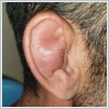
AbstractAuricular hematoma is a condition that involves blood accumulation between auricular perichondrium and underlying cartilage tissue, resulting in long lasting disfigurement of ear. Most common cause for auricular hematoma is traumatic etiology. Spontaneous auricular hematoma is quite rare. Behçet disease, a vasculitic disorder, is unique among vasculitic diseases for ability to affect all vessels. Vasculitis may lead to bleeding complications. This study presents first case in literature with spontaneous auricular hematoma in Behçet disease.IntroductionDirect trauma to the auricle may cause seperation of auricular perichondrium from underlying cartilage tissue and blood accumulation between these tissues, a condition known as auricular hematoma[1]. Cartilage is dependent on perichondrium for vascularity, thus auricular hematoma may cause cartilage deformities and long lasting disfigurement of ear[2]. Trauma is the main reason beneath auricular hematoma; but infectious causes, bleeding disorders and rheumatologic entities also should be kept in mind. Spontaneous non-traumatic auricular hematomas are quite rare. Treatment generally involves incision, drainage and suturation of ear with different materials such as ear dressings or buttons. Behçet disease is a vasculitic disease described by Professor Hulusi Behcet in 1937. This disorder is commonly known with a particular triad: recurrent oral aphtous ulcers, genital ulcers and uveitis. Among vasculitic disorders, Behcet disease is unique for ability to affect all vessels with no regard to size. Vasculitis may cause many complications, involving different hematomas. We present you a case, first example of spontaneous auricular hematoma in Behçet disease in literature. Case ReportA 33-year-old male presented to our clinic with the complaint of sudden swelling of right ear. According to the patient, the swelling started a couple hours ago and he denied any recent trauma, bleeding disorder, anticoagulant use or insect bite. Only remarkable thing in history is that patient had Behçet disease known for 8 years. Laboratory tests had shown normal complete blood count, electrolytes and coagulation tests. Physical examination revealed a bulging, fluctuant swelling and redness in right auricle; consistent with auricular hematoma (Figure 1).
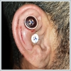
Urgent surgical intervention began with infiltration anesthesia with %2 lidocaine with adrenaline. Then, an incision was made with a #15 no. scalpel between helix and anti-helical fold. Blood accumulate was gently aspirated with Frazier suction. Two buttons were sewed anteriorly to auricle to prevent re-accumulation (Figure 2).
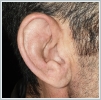
At fifth day, sewn buttons are removed. After 3 months, patient is complication free with satisfactory healing (Figure 3).
DiscussionBehçet disease is a chronic relapsing and remitting vasculitis of unknown aetiology and unique for ability to affect all vessels with no regard to size. Incidence is severely high in silk road countries, regarding to genetical predisposition. Most commonly known genetical factor in pathogenesis of Behcet disease is self-antigenic role for Human leucocyte antigen-B51[3]. Most famous symptom triad of Behcet disease consists of recurrent oral aftous lesions, ocular disease and genital ulcerations. In addition to that, many other organ systems may be involved in course of disease, such as central nervous, gastrointestinal, cardiovascular, pulmonary systems and skin[4]. Vasculitis in Behcet disease is able to cause thrombosis and bleeding complications. There are some reports in literature for hemorrhagic complications of Behçet disease such as spontaneous retroperitoneal, recurrent intracranial and pulmonary haemorrhages[5-7]. Most common otological manifestations of Behcet disease are hearing loss and vertigo due to vasculitic inner ear involvement. These otological findings may be the first symptoms of a undiagnosed Behcet disease and patient should be evaluated as a whole[4]. As far as we know, auricular hematoma are not a common otologic manifestation of Behcet disease. Our patient with Behçet disease had spontaneous auricular hematoma, a pretty rare clinical condition, first in English literature as far as we know. Treatment for auricular hematoma is simple incision, drainage and suturation[2]. Suturation aims to provide adequate compression and preventing re-accumulation. Shakeel et al. defined mattress suturation with absorbable sutures is the best way to prevent re-accumulation of hematoma[8]. Some authors recommend that suturation should involve hard materials such as buttons[9]. In delayed cases with deformed ears, newly and irregularly developed cartilage may need to be excised. Most common complications of surgical intervention are auricular cellulitis, permanent ear deformities due to re-accumulation and suture abscesses[8]. We lacked absorbable suture materials in our clinic, thus we used suturation with buttons instead of mattress sutures after incision and drainage in our patient. No complications had been seen so far in our 3 months of follow up. Most common cause for auricular hematoma is traumatic etiology. Spontaneous auricular hematoma is quite rare. As far as we know, there’s no patient in literature with spontaneous auricular hematoma in Behçet disease. It may be hard to link Behcet disease to spontaneous auricular haematoma in only one case, but vasculitis in Behcet disease is known for many spontaneous hemorrhagic complications. In literature, there are patients with undiagnosed Behcet disease, only to be known after some hemorrhagic complications, such as spontaneous retroperitoneal hemorrhage. We aimed to draw attention to Behçet disease, a rheumatologic disorder that is common in our country, likewise other silkroad regions, could be a unknown cause beneath spontaneous auricular hematoma. In Turkey and other silkroad countries with high Behcet disease incidence; patients with spontaneous auricular hematoma should also be evaluated for Behcet disease. References
|
|||||||
| Keywords : spontan , auriküler , hematom , Behçet hastalığı | |||||||
|


