

Authors
|
|||||
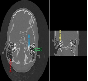
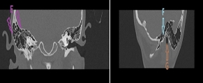
AbstractMastoid hyperpneumatization is an uncommon mastoid bone pathology, mostly seen in adult males, develops spontaneously and is generally noticed incidentally. In this study, 3 cases of mastoid hyperpneumatization detected radiologically are presented in light of current literature.IntroductionThe relationship between the development of mastoid pneumatization and middle ear diseases is known. Discussions on the factors affecting the development of pneumatization are gathered in two main views: genetic and environmental theory. Patients most often refer to the physician with complaints of swelling in the mastoid region. Other complaints are headache, moderate conductive hearing loss, ringing in the ear, neck pain, numbness in the left arm, and hypoglossal palsy. Although the most common complications are extradural and extracranial pneumatocele, foramen magnum emphysema can also be seen [1]. In this study, 3 cases of mastoid hyperpneumatization detected radiologically are presented in light of current literature. Case ReportWithin 3 months, 3 patients applied to our outpatient clinic with a complaint of swelling behind the ear. Two of the patients were boy and girl aged 9 and 10 years old, respectively. One patient was a 23-year-old female. Bilateral mastoid bone hypertrophy was observed in the physical examination of three patients. On examination, bilateral tympanic membranes were natural in all patients. Computer tomography (CT) was requested for patients with no suspected abnormality in tympanogram and audiometric tests (Figure 1,2).
Ventilation tube was recommended to patients with bilateral mastoid hyperpneumatization in CT, but all three patients refused surgical procedures. The patients were advised to change their nasal cleansing habits. DiscussionEasily accessing imaging methods such as CT and MRI and an increasing number of patients screened with these imaging methods often cause physicians to encounter more pathological conditions such as mastoid hyperpneumatization. The process of the pneumatization of the temporal bone starts in the last weeks before birth with a reduction of the embryonic mesenchyme in the antrum. This process progresses through infancy, childhood, and puberty until the last part of the petrous apex is pneumatized. Large pneumatization of the temporal squama, extending behind the sigmoid sinus, is considered to be a normal variant. Pneumatization of temporal bone areas such as petrous apex, mastoid, squamomastoid bone, and perilabyrinth is considered physiological in adults [2]. There are very few cases in the literature associated with mastoid hyperpneumatization, mostly detected by trauma by chance. When it is symptomatic, it usually presents with symptoms of otolaryngology such as tinnitus and vertigo [3-5]. Many hypotheses have been proposed about its occurrence, including developmental anomaly, the pressure increase in the middle and inner ear. Mastoid pneumatization begins after birth, and in the first years of life, mastoid cells are formed in different forms. A dorsal extension of the cavum tympani forms the antrum mastoideum. It connects the epipharynx and middle ear, which develops from the proximal of the first pharyngeal pouch, and this connection causes inflammation of the middle ear and mastoid bone after infections of the upper respiratory tract [6]. Martin et al. [7] attributed the attenuation of the atlantooccipital and mastoid cells to the eustachian tube working like a valve and creating a driving force. On the other hand, some authors attributed this to the embryonal developmental anomaly. Decrease of mesenchymal tissue causes incomplete fusion of occipiomastoidal synchondrosis and mastoid hyperpneumonitization [3]. Patients should be aware that even a small trauma to the occipitomastoid region can cause a very thin outer laminate to break and to cause any complications. These complications are the most common extracranial pneumotocele, but can also cause extradural pneumotocele and pheromenal magnum emphysema. On the other hand, if the eustachian tube is working well and the bone remains intact, the cases described in hyperpneumatization remain asymptomatic. Although different surgical methods such as mastoidectomy, canaloplasty, mastoid explanation, and tube insertion have been tried to date. Some authors also suggest only follow up. References
|
|||||
| Keywords : Hiperpnömatizasyon , mastoid , temporal kemik , bilgisayarlı tomografi | |||||

|