

Authors
|
|||
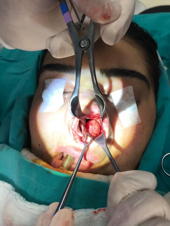
AbstractNasoalveolar cysts are extremely rare, nonodontogenic soft tissue lesions. It is often diagnosed at an early stage because it often causes cosmetic problems. In this study, a case who was operated for septorhinoplasty and whose nasoalveolar cyst causing nasal deformity was detected incidentally peroperatively was presented in the light of current literature.IntroductionNasoalveolar cysts, which originate from epithelial remnants of the nasolacrimal duct, are nonodontogenic soft tissue lesions of the upper jaw [1]. They usually cause painless swelling and rarely nasal congestion unless they are infected [2]. Clinical and histopathological findings give the diagnosis [3]. On medical exams; they appear as well-restricted, mobile, fluctuating masses. They are best examined by palpation at the base of one hand on the nose and the other hand on the gingivolabial sulcus [4]. The treatment aims at correcting the deformity, removing the nasal obstruction, and treating if there is an infection. In this study, a case who had septorhinoplasty and whose nasoalveolar cyst causing nasal deformity was observed incidentally perioperatively was presented in the light of current literature knowledge. Case ReportA 36-year-old female patient came to our clinic with the complaint of prolonged and increasing nasal congestion and deformity in the nose. A medical exam showed hypertrophy in both lower turbinates, deviation to the right in the septum, nasal hump, and a shift to the left in the nasal axis. Other examinations were natural (Figure 1). There was no prominent feature in the patient's history, except for allergic rhinitis. Since another pathological finding, other than turbinate hypertrophy, was not observed, no radiologic imaging was requested. Septorhinoplasty and concha reduction operation was planned for the patient. During the surgery, the cystic formation of approximately 3x3 cm in size, with a smooth surface was observed under the left lower turbinate that caused the turbinate leaning medially and the left nasal bone laterally (Figure 2). The cyst was removed with a sublabial approach. Then, the operation of septorhinoplasty was completed and the procedure was ended without complications. In pathologic examination, light microscopy of the cystic wall showed a connective tissue with few cells, lined with a flattened squamous epithelium. Microscopically, the cyst wall showed foci of chronic inflammatory cells, and the wall was lined with two layers of squamous epithelium. The lession was verified to be nasoalveolar cysts by histopathological examination. No recurrence was observed in the 6-month follow-up of the patient. DiscussionHistologically, it is a common thought that the cause of the nasoalveolar cyst is the "fissural cyst", which is caused by a defect in the fusion of the medial nasal wall, lateral nasal wall and the maxillary process in the intrauterine first month [5]. As for histopathological evaluation, Su et al. in their scanning electron microscopic exam revealed that cilia observed in the light microscope were numerous short globular or irregular microvilli which probably resulted from lack of stimulation by air. [6]. Allard encountered only seven cases of nasoalveolar cysts in a study in which they examined 8000 cystic lesions of the oral cavity over 10 years [7]. This number is probably lower since the cyst was not noticed without infection or creating facial deformity [8]. The nasoalveolar cyst was first described by Zuckerkandl in 1882 [1]. For the nasoalveolar cyst; although names such as nasoalveolar cyst, mucoid cyst, subalar cyst, nasal vestibular cyst, nasal wing cyst are used, the most common use in the literature is the nasoalveolar cyst [9]. Although it can be seen at any age, it is more common in 4-5 minutes [10,11]. It is generally unilateral and more common on the left and in women and may be bilateral at 11.2% [2]. In our case, the cyst was located on the left side following the literature and our patient was female. On physical examination, a mobile smooth cystic mass in the nasoalveolar groove can be palpated [12]. In our case, there were no other findings other than nasal deformation and turbinate hypertrophy. There is an increased risk of infection in nasoalveolar cysts due to their proximity to the mouth and nose. In his study, Kuriloff reported that half of his patients developed an infection [13]. As an initial symptom, cyst infection and related complaints are seen in 30% of patients [8]. No signs of infection were detected in our patient during diagnosis, treatment, and follow-up. Although the diagnosis of nasoalveolar cyst is clinical, rarely seen cyst usually results in the use of imaging methods in patients [14]. They do not destroy neighboring bone structures, but they may lead to limited sclerotic density increases in the cortical-subcortical area, which can be monitored in small amounts of deformation and conventional radiological examinations [3]. In the study of Raphael N. Aquilino et al., There were no pathological findings in plain radiographs such as panoramic radiography, and magnetic resonance [MR] changes were determined according to the cyst content. In diagnosis, CT was seen as an adequate option because it provides sufficient information about the localization of the cyst, its structure, especially the relationship of the cyst with the surrounding tissues and bone erosion [8,14]. Since the cyst content in MR is seen more clearly than CT, it should be preferred after CT in suspicious lesions [3]. No preoperative imaging was requested in our case since there was no external swelling and septorhinoplasty was planned for the patient. Treatment of the nasoalveolar cyst is surgical excision with a sublabial approach [2,3,15]. Recurrence after treatment has never been reported, and only one case with malignancy has been reported [3]. In the differential diagnosis of the disease, neoplastic, developmental, and odontogenic lesions that may be soft, benign, and painless should be considered. Nasoplatin duct cyst or incisive duct cyst can often be confused. Periapical inflammatory lesions such as granulation, cyst, and an abscess can erode the bone and mix with this lesion. One of the developmental lesions progressing aggressively, keratocyst can also be encountered with bone erosion. Nonodontogenic epidermoid or epidermal inclusion cysts can be in a similar clinical picture. Other diseases that should be considered should be the nasal floor furuncle, facial cellulitis, acute maxillary sinusitis [8]. In conclusion, the nasoalveolar cyst is one of the rare maxillofacial cysts that may be encountered by otolaryngologists. Although the diagnosis can be made clinically, additional imaging methods are required for differential diagnosis. Unlike other case reports in the literature, the feature of our case is; This is the presence of cystic formation in the patient, which does not show any obvious findings from the outside, but causes expulsion in the nasal bone and repulsion in the turbinate, which makes breathing difficult and incidentally detected during septorhinoplasty. Septorhinoplasty has become a frequently used procedure in otolaryngology practice today. Generally, physicians do not need preoperative imaging. Our aim with this case is to report that this antithesis, which is rare but may cause nasal deformation, should be kept in mind and that it will be beneficial not to encounter surprise during operation in case of suspicion in patients with asymmetric concha hypertrophy. References
|
|||
| Keywords : Septorinoplasti , burun kemiği , nazoalveoler kist | |||
|