|
CAVERNOUS HEMANGIOMA OF THE MIDDLE TURBINATE DIAGNOSED AS CONCHA BULLOSA RADIOLOGICALLY
CAVERNOUS HEMANGIOMA OF THE MIDDLE TURBINATE DIAGNOSED AS CONCHA BULLOSA RADIOLOGICALLY
Articles >
Rhinology
Submitted : 10.05.2020
Accepted : 08.07.2020
Published: 08.07.2020
|
|
Abstract
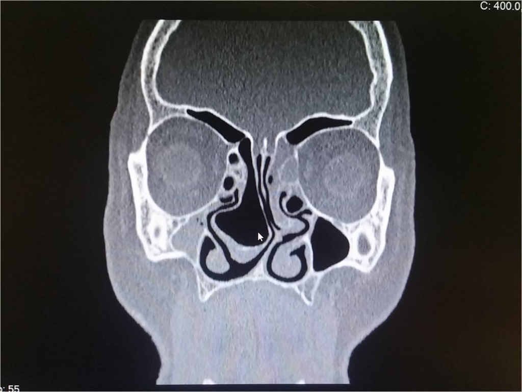
The nasal cavity is an uncommon site for a cavernous hemangioma. Cavernous hemangiomas of middle turbinates are very rare. Pneumatization of the middle turbinate, also known as concha bullosa, is a common anatomical variation of the sinonasal region. We describe a case of a cavernous hemangioma of the nasal cavity with a prediagnosis of concha bullosa of the middle turbinate according to computed tomography (CT) findings. Paranasal CT studies showed pneumatization of the middle concha. Given the prediagnosis, partial resection of the middle turbinate was performed instead of complete resection. The patient was followed up for 2 years. There was no enlargement of the middle turbinate after surgery. Three interesting aspects of the present case were the rarity of the site of the hemangioma, nonspecific symptoms, and radiological findings, which pointed to a different pathology i.e., concha bullosa. To our knowledge, this is the first reported case in the English literature of cavernous hemangioma with a prediagnosis of concha bullosa.
|
|
Keywords:
cavernous hemangioma
, middle turbinate
, concha bullosa
|
Özet
Nazal kavite kavernöz hemanjiyom için nadir görülen bir bölgedir. Orta konkaların kavernöz hemanjiyomları çok nadirdir. Konka bulloza olarak da bilinen orta konkanın pnömatizasyonu, sinonazal bölgenin genel anatomik bir varyasyonudur. Bu yazıda bilgisayarlı tomografi (BT) bulgularına göre, orta konka bulloza ön tanısı konulan bir kavernöz hemanjiyom olgusu sunulmuştur. Paranazal BT'de orta konkadaki pnömatizasyon görülmektedir. Ön tanıya dayanarak, tam rezeksiyon yerine orta konkanın kısmi rezeksiyonu yapılmıştır. Hasta 2 yıl boyunca takip edilmiş, ameliyat sonrası orta konkada genişleme olmadığı görülmüştür. Mevcut vakanın üç ilginç yönü, hemanjiyomun bulunduğu yerin nadirliği, spesifik olmayan semptomlar ve farklı bir patolojiye işaret eden radyolojik bulgular, yani konka bulloza ön tanısıdır. Bildiğimiz kadarıyla, İngilizce literatürde konka bulloza ön tanısı konulan ilk kavernöz hemanjiyom olgusudur.
|
|
Anahtar kelimeler:
kavernöz hemanjiom
, orta konka
, konka bulloza
|
|
|
|