

|
|||||
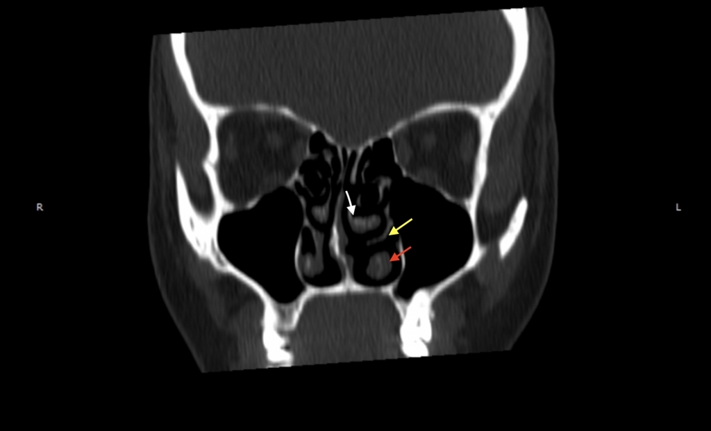
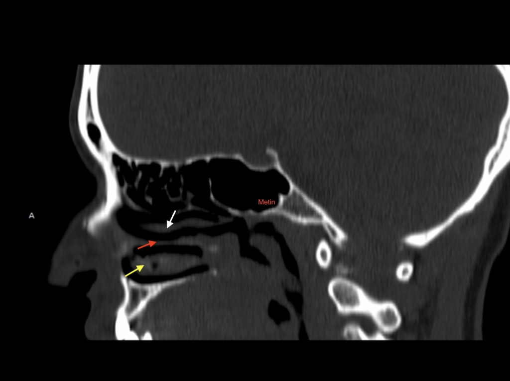
AbstractThe anatomy and embryology of the nasal cavity and paranasal sinuses are quite complex. Nasal turbinates are important anatomical structures that extend from the lateral nasal wall to the nasal cavity. After the widespread use of endoscopes and computed tomography, many variations of these structures have been described. The secondary middle turbinate is a one of the rare variations of the nasal cavity. It is a soft tissue-covered bony prominence arising from the lateral nasal wall. It usually causes sinusitis by closing the osteomeatal complex. We present the case of a female patient diagnosed with this rare anatomical variation by means of computed tomography. On examination, a mass thought to be a nasal polyp was detected in the left nasal cavity. The computed tomography of the paranasal sinuses was done which showed a bony structure attached to the lateral wall of the middle meatus and covered by a soft tissue. It diagnosed as secondary middle turbinate and then the patient was treated with surgical intervention. Accurate identification of the anatomical variations of the turbinates will help to determine the correct diagnosis and treatment and to prevent possible complications during endoscopic procedures.IntroductionThe three nasal turbinates, defined as the superior, middle, and inferior turbinate, extend from the lateral nasal wall to the nasal cavity [1]. During the complex embryological process of the nasal turbinates, anatomical variations may occur at more than one period [2]. The middle turbinate and superior turbinate develop embryologically from the second and third ethmoturbinal. While these are part of the ethmoid bone, the inferior turbinate develops as a separate bone from the maxilloturbinal. The uncinate process (UP) originates from the first ethmoturbinal [3,4]. Concha bullosa (CB), the pneumatization of the turbinate, is the most widespread anatomical variation of the turbinates. The others include the duplicate turbinate, paradoxically curved middle turbinate, turbinate, secondary middle turbinate (SMT) and sagittally clefted middle turbinate [5-8]. These variations may cause inflammatory pathologies. Furthermore, it is important surgically as the middle turbinate is a significant marker in endoscopic sinus surgery. SMT is an uncommon anatomical variation that originates from the lateral wall of the middle meatus and protrudes mostly superomedially, therefore it can be easily confused with a nasal polyp, osteoma, or sinonasal tumor. In this article, we present a female patient in whom this rare anatomical variation was detected incidentally by computed tomography. Case Report40-year-old female patient, presented with several year- history of nasal obstruction. In the endoscopic examination performed in the outpatient clinic, the nasal septum was deviated to the left and a mass thought to be a nasal polyp was detected in the left nasal cavity. Computed tomography of paranasal sinuses confirmed the deviated nasal septum to the left; however, it also demonstrated a bony structure attached to the lateral wall of the middle meatus and covered by a soft tissue. This finding was compatible with a diagnosis of a secondary middle turbinate (SMT), which was shown initially in endoscopic examination as a left nasal polyp (Figure1-2).
She had no history of previous nose surgery. After diagnosis of this sinonasal anatomic variation, the patient underwent septoplasty, and bilateral inferior turbinates were ablated with radiofrequency. DiscussionThe nasal turbinates extend from the lateral nasal wall into the nasal cavity. The embryological development of the turbinates is a quite complex process. The progenitor structures of the nasal turbinates, ethmoturbinal and maxilloturbinal, emerge between the eighth and tenth weeks of fetal life. While the inferior nasal turbinate develops from the maxilloturbinal, the UP, middle turbinate, superior turbinate and, if present, the supreme turbinate originate from the ethmoturbinal [10]. During this process, anatomic variations may occur at multiple periods. The most common anatomical variation is the pneumatization of the middle turbinate [9]. The other variations of turbinates include ethmoidal bulla, agger nasi cells, Haller cells, paradoxical middle turbinate, pneumatized uncinate process, deviated uncinate process, accessory middle turbinate, middle turbinate, and bifid inferior turbinate. [10] Khanobthamchai et al. defined SMT as a protruding bone originating from the lateral nasal wall just below the basal lamella [11]. Aksungur argued that the SMT may be an additional turbinate originating from part of the frontal ridge [5]. The incidence of SMT is approximately in the range of 0.8 to 14.3% [4,5]. SMT can be seen unilaterally or bilaterally so it may be confused with a simple nasal polyp or a nasal tumor during endoscopic examination [6]. Ark et al. presented a case of unilateral SMT with sinusitis [12]. In our case SMT was detected unilaterally, too. Osteomeatal complex obstruction may not be seen in SMT cases with a downward and medial curvature. However, in the study of Apaydın et al., SMT curves inferiorly and medially towards the middle meatus and narrows the underlying hiatus semilunaris slightly [6]. In patients without maxillary sinus infection clinically and radiologically, the patient can be followed up without any treatment. ConclusionAs a result, other authors have concluded that SMT is generally a bilateral anatomical variation. However, in this case we found that SMT can occur unilaterally, emphasizing a variation that should be kept in mind, it may possibly be confused with other conditions such as nasal polyps and tumors. Informed ConsentWritten informed consent was obtained from patient.References
|
|||||
| Keywords : Nazal konka , anatomik varyasyon , ikincil orta konka , kulak burun boğaz. | |||||

|