

Authors
|
|||||
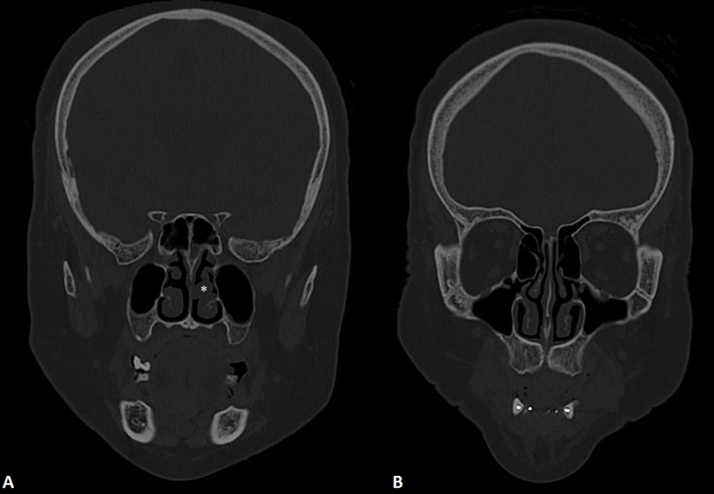
AbstractChoanal polyps are defined as benign, inflammatory, solitary soft tissue lesions and usually originate from the maxillary sinus. The choanal polyps can originate from unusuallocations such as the sphenoid sinus, ethmoid sinus, nasal septum, hard and soft palate. Choanal polyp arising from middle turbinate is an extremely rare entity. In this report, a case of solitary choanal polyp that originate from middle turbinate and successfully removed with endoscopic surgery is presented. IntroductionChoanal polyps (CPs) are benign, solitary soft tissue lesions that usually arise from the maxillary sinus and extend toward the nasopharynx [1]. Choanal polyps can originate from unusual locations such as the sphenoid sinus, ethmoid sinus, nasal septum, hard and soft palate [2]. The origin from the middle turbinate with extension to the choana is very rare. To the best of our knowledge, only four cases have been reported in the English literature [2-5]. Antrochoanal polyps (ACPs) arise from inflamed mucosa of the maxillary sinus and mostly have two components; a cystic part and a solid part. ACPs pass through the maxillary ostia into the middle meatus, with extension to the nasopharynx or oropharynx. The cystic part fills the maxillary sinus and is attached to the solid part, which fills the nasal cavity with a pedicle [1]. Herein, we present a rare case of CP, which arised from the lateral side of middle turbinate, and discuss the radiological and surgical findings. Case ReportA 40-year-old female patient presented to our department with a left-sided nasal obstruction. There was no history of nasal allergy. She had hypertension and diabetes mellitus. Endoscopic nasal examination revealed a choanal polyp (CP), arising from the lateral side of middle turbinate and extending to the choana. Computed tomography scan demonstrated a polyp that arised from lateral side of the middle turbinate, extending posteriorly towards the choana and filling the nasopharynx. Left middle meatus and ostiomeatal unit were clear (Fig. 1A and B).
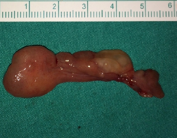
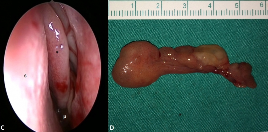
Written informed consent was obtained from the patient. Endoscopic endonasal surgery was performed to patient under local anaesthesia with sedation. The polyp’s pedicle was followed to the origin on the middle meatus and the polyp was completely removed (Fig. 2C). The pathological inspection revealed a single, smooth surfaced polypoid tissue that measured 6x2x1 cm (Fig. 2D). The histopathological diagnosis was inflammatory polyp. There is no recurrence or complication in 12 months follow-up on nasal examination.
DiscussionAlthough there are different types of choanal polyps, most of them arise from maxillary sinus and are called “antrochoanal polyps”. ACPs arises from inflamed mucosa of the maxillary sinus and mostly have two components; a cystic part and a solid part. [6] They almost always occur in children and young adults [1]. CP can originate from the ethmoid sinus, sphenoid sinus, inferior nasal concha, nasal septum in rare cases [4,7,8,9]. A choanal polyp arising from the middle turbinate with an extension to choanae is very rare entity. Only 4 cases have been reported in the English literature [2-5]. In three of them the origin was from the medial side of the middle turbinate and one of them originated from inferior side of the middle turbinate. In our case, CP is originating from lateral side of middle turbinate. Although varying in location, CPs present with same symptoms and histological findings. As in our case, the most common symptom is nasal obstruction. Unlike ACPs, CPs are not seen commonly in children [2]. Including our case, all of the reported middle turbinate originating choanal polyp cases were adults. Diagnosis of a CP is made by nasal endoscopic examination and imaging modalities. Computed tomography is very helpful in making diagnosis, determining the site of origin and the extent of the polyp. Differentiation of CP with ACP is important for surgical planning. ACPs develop between the middle turbinate and lateral wall of the nasal cavity, on the other hand the CPs that originate from middle turbinate fill the space between the middle turbinate and septum. In addition, CPs can occasionally be part of nasal polyposis, therefore it must be distinguished from generalized nasal polyps [4]. The differential diagnosis of CP should include angiofibroma, haemangioma, lymphoma, retention cyst, mucocele, mucopyocele, inverted papilloma, turbinate hypertrophy, adenoid hypertrophy, Tornwaldt’s cyst and olfactory neuroblastoma. In particular, inverted papilloma should be considered if there is an unusual site of origin of a CP [4]. Endoscopic sinus surgery is recommended treatment and provides low recurrence rate [2]. A careful endoscopic resection of the polyp and the mucosa of its point of origin, must be performed to prevent recurrence [8] In conclusion, CP can originate from unusual locations and it should be considered in the differential diagnosis of patients presenting with nasal obstruction. We recommend endoscopic surgery under local anesthesia for choanal polyps originating from the middle turbinate. References
Presented atThis case report presented in 36th Turkish National Congress of Otorhinolaryngology Head and Neck Surgery. |
|||||
| Keywords : Koanal polip , Orta konka , Endoskopik cerrahi , Burun tıkanıklığı | |||||
|