

Authors
|
|||||
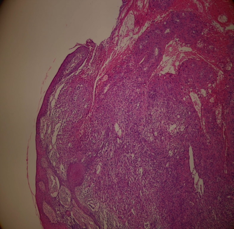
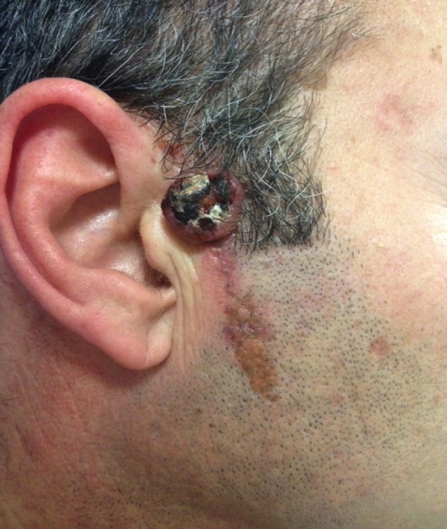
AbstractNevus sebaceous is a benign hamartoma of the skin, characterized by hyperplasia of the epidermis. Hovewer mostly benign tumors occur on nevus sebaseus; It was reported that malignant carcinomas can also occur.Basososqumaous cell carcinoma (BSC) is a rare carcinoma. It is mostly seen in male and in head and neck region. This is the first paper in the literature that reports Basosquamous carcinoma (BSC) arising on a linear nevus sebaseous which has not been reported in the previous studies. IntroductionNevus sebaceous is also called nevus sebaceous of Jadassohn or organoid nevus. It is a benign hamartoma of the skin. Nevus sebaceous is characterized by hyperplasia of the epidermis, immature hair follicles, and sebaceous and apocrine glands [1]. Nevus sebaceous usually occurs in the head and neck region and is usually clinically apparent at birth. It presents as a well-demarcated, skin-colored to yellowish alopecic patch. Proportional enlargement with age is the rule and at puberty the lesion typically becomes more yellowish and cerebriform. The secondary tumors arising within nevus sebaceous and the risk of malignant neoplasm have been controversial [2]. Herein we report a case of Basosquamous Carcinoma (BSC) arising on a linear nevus sebaceous, which has not been reported in previous studies. Case ReportA 52-year-old man was admitted our clinic with a dome shaped bump on a previously existed nevus on his face, which had grown in the past three months. On dermatological examination, a 0.5x7 cm, bright yellowish verrucous plaque was located on the right preauricular region, arising from the mandibular angle and extending into the hairline on the temporal area in a linear pattern, which has been present since birth. There was a 1.3 cmx1 cm nodule on the linear lesion with a black crust and an ulcer in the center (Figure 1). The otorhinolaryngologic examination was normal. The nodular lesion was totally excised with a 1 cm margin because of the prediagnoses of basal cell, squamous cell carcinoma and keratoacanthoma. The histopathological examination revealed BSC. The surgical margins were negative. Tumor cells had large vesicular nuclei, prominent nucleoli and narrow cytoplasm. The hyperchromatic nuclei in adjacent areas, narrower cytoplasm of tumor epithelial islands form has attracted the attention associated with tumor formation of basal layer (Figure 2). He was diagnosed with BSC of the sebaceous nevus with these findings. The common blood count and biochemical laboratory tests were within normal ranges. There were no pathologic lymph nodes on the cervical ultrasonographic examination, and PETCT was normal. The chest x-ray radiographic examination was normal. After one year, there were no signs of recurrence in his follow-up.
DiscussionVarious adnexal tumors can occur on sebaceous nevi, especially in adults. However, mostly benign tumors occur on sebaceous nevi. It was reported that basal cell carcinoma, squamous cell carcinoma, apocrine carcinoma, sebaceous carcinoma, adenomyoepithelioma, and microcystic adnexal carcinoma can develop on sebaceous nevi [3]. BSC is a rare epithelial neoplasm with features of both basal cell carcinoma (BCC) and squamous cell carcinoma (SCC). BSC has more aggressive behavior and has higher risks for recurrence and metastases [4,5]. Clinically, BSC is mostly seen on the head and neck, and has a significant predominance in males. Most of the BSC were located in the head and neck, with a higher rate seen on the nose (33.1%). The auricular region and periocular region are also the most commonly affected sites [6,7]. The patient in the present case had a tumor located in the preauricular region, and had a history of sun exposure due to his job. BSC has a nonspecific clinical presentation and the diagnosis is made only after biopsy. The best treatment for BSC is total excision with wider surgical margins because BSC has an infiltrative growth pattern. Because of its aggressive behavior, long-term follow-up for the detection of local recurrence and distant metastatic spread is recommended. Conclusion It should be kept in mind that BSC can be one of the malignant tumors that develops on the sebaceous nevus.
References
Presented at25. Ulusal Dermatoloji Kongresi 2014, Antalya, 21 Ekim 2014 - Poster Sunumu |
|||||
| Keywords : Bazoskuamöz Hücreli Karsinom , Nevus Sebaseus , Cilt Kanserleri | |||||
|