Abstract
Foreign bodies are frequently encountered in otorhinolaryngology. Adult patients usually encounter accidents involving foreign bodies. In this case report, we present a patient of which an iron piece was accidentally inserted between the left eye and the left side of the nose while cutting marble.
Introduction
Foreign bodies are frequently encountered in otorhinolaryngology. Patients, in general, are at increased risk in adults with mental retardation, as well as in the pediatric age group. Adult patients usually encounter accidents involving foreign bodies. The success of taking foreign bodies depends on several factors such as foreign body location, cystic material, comprehensible (soft and irregularly edged) or incomprehensible (hard and round), physician's skill and patient co-operation [1]. In this case report, we present a patient of which an iron piece was accidentally inserted between the left eye and the left side of the nose while cutting marble.
Case Report
A thirty-one-year-old male patient stabbed a foreign body (iron piece) between the left eye and the left side of the nose while cutting marble with a cuuting tool. Patient who had no complaints of loss of consciousness and bleeding applied to our emergency department after accident in 30 minutes.
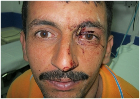
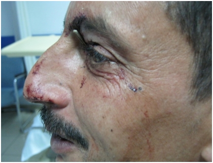
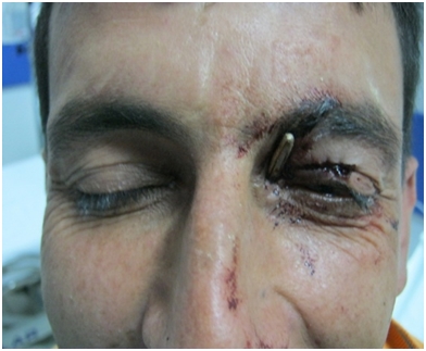
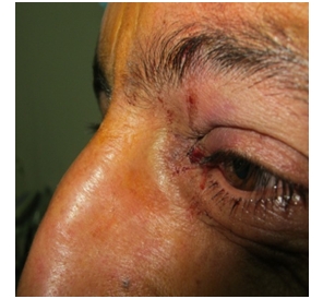
The patient was evaluated in emergency unit. There was an iron piece in the medial part of the left eye that was seen about 2 cm outside the epicentus line (Figure 1,3,5,6). As a result of consultation of ophtalmologist, the eye movements and vision of the patient were evaluated natural. A foreign body was not seen in the endoscopic examination of the patient, but the patient felt severe pain when moving the left middle concha.
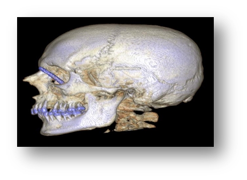
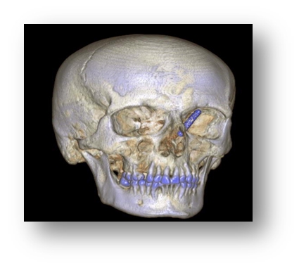
In the facial CT, there was a linear metallic foreign body that started from the medial cantus level on the left side and proceeded to the posteroinferomedial region and partially traumatized the anterior ethmoidal cells and extended to the left osteomeatal complex level (Figures 2,4,7).
The patient was taken to the operating room immediately. Foreign body was removed under local anesthesia (Figure 8). Foreign body was found to be about 4 cm in size. No complications were seen after removal. Visual and eye movements were evaluated natural (Figures 9). Patient was discharged one day later.
Discussion
Foreign bodies may be retained in the body through different mechanisms, including work accidents ingestion, placement in bodily orifices, and surgical errors. In the United States in 1999, there were 8.2 million emergency department visits for open wounds with foreign bodies [2] People who work in occupations such as carpentry and the garment industry are at increased risk of insertion with nails, pins or different metal or wooden parts. These injuries are more common in children or adults with mental or physical impairment, which may result in behavior or lack of control that increases risk [3].
Although these injuries may seem minor, wounds with neglected foreign bodies are a common cause ofinfections and malpractice claims [4]. Any wound that penetrates the skin should be evaluated to determine if exploration for foreign bodies is needed. The mechanism of injury is important in evaluating for foreign bodies. Bite injuries may include teeth, and punches to the face may include tooth fragments in the punching hand. Broken objects causing wounds may leave embedded fragments [5].
Being adequately prepared for the removal of foreign bodies increases success rates and avoids complications. Wound exploration is aided by optimal lighting, magnification, and adequate hemostasis. The wound and gloves should be cleansed before removal is attempted. Anesthesia is necessary for deeply embedded fishhooks, larger splinters, or wound exploration. Local infiltration or digital block can be used depending on the location of the wound. For children, it may be beneficial to use topical anesthetics [6]. Compared with a eutectic mixture of local anesthetics, 4% liposomal lidocaine has a shorter application time and longer duration of action with good pain control [7]. The most important way to avoid infection is to completely remove the foreign body. After removal, if the wound is large enough, it can be irrigated with drinkable tap water [8,9]. In a puncture wound, injecting saline under pressure may drive contaminants further into tissue and should be avoided. Antiseptic agents such as hydrogen peroxide, chlorhexidine, and povidone iodine should not be used because they are toxic to tissue and slow the healing process [10,11]. We performed local anesthesia for removal. No complication had occured.
Although infection is the most common complication, with rates ranging from 1.1 to 12 percent, the use of prophylactic antibiotics is not typically recommended in nonbite wounds [12]. Antibiotics may reduce the rate of infection after bites by humans and after bites on the hand [13].We prescribed antibiotic for patient even it was a clean wound.
References
- Steven W. Heim, MD, MSPH, and Karen L. Maughan, MD University of Virginia School of Medicine, Charlottesville, Virginia,Foreign Bodies in the Ear, Nose, and Throat, Am Fam Physician 2007;76:1185-9
- National Center for Health Statistics. Emergency department visits. Accessed April 23, 2007, at: http://www.cdc.gov/nchs/fastats/ervisits.htm
- Halaas GW, MD, MBA, University of Minnesota Medical School, Minneapolis, Minnesota, Management of Foreign Bodies in the Skin, Am Fam Physician, 2007; Volume 76, Number 5
- Vukmir RB. Medical malpractice: managing the risk. Med Law 2004; 23:495-513.
- Capellan O, Hollander JE. Management of lacerations in the emergency department. Emerg Med Clin North Am 2003;21:205-31.
- Chen BK, Eichenfield LF. Pediatric anesthesia in dermatologic surgery: when hand-holding is not enough. Dermatol Surg 2001;27:1010-8.
- Eichenfield LF, Funk A, Fallon-Friedlander S, Cunningham BB. A clinical study to evaluate the efficacy of ELA-Max (4% liposomal lidocaine) as compared with eutectic mixture of local anesthetics cream for pain reduction of venipuncture in children. Pediatrics 2002;109:1093-9.
- Valente JH, Forti RJ, Freundlich LF, Zandieh SO, Crain EF. Wound irrigation in children: saline solution or tap water? Ann Emerg Med 2003;41:609-16.
- Fernandez R, Griffiths R, Ussia C. Water for wound cleansing. Cochrane Database Syst Rev 2002;(4):CD003861.
- Krasner D. AHCPR Clinical practice guideline number 15, treatment of pressure ulcers: a pragmatist’s critique for wound care providers. Ostomy Wound Manage 1995;41(7A suppl):97S-101S. Accessed April 23, 2007, at: http://www.ncbi.nlm.nih.gov/books/bv.fcgi?rid=hstat2.chapter.5124.
- Oberg MS, Lindsey D. Do not put hydrogen peroxide or povidone iodine into wounds! Am J Dis Child 1987;141:27-8.
- Cummings P, Del Beccaro MA. Antibiotics to prevent infection of simple wounds: a meta-analysis of randomized studies. Am J Emerg Med 1995;13:396-400.
- Medeiros I, Saconato H. Antibiotic prophylaxis for mammalian bites. Cochrane Database Syst Rev 2001;(2):CD001738
|