

|
|||||||||||
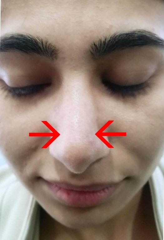
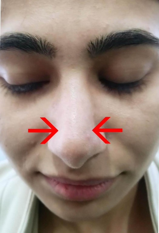
AbstractSynkinesis is the contraction of involuntary muscle group synchronously to the contraction of voluntary muscle groups. Simultaneous contraction of oculonasal synkinesis, orbicularis oculi, and compressor narium minor muscle. Oculonasal synkinesis is a rare phenomenon.In a 18-year-old female patient who had undergone open technical septorhinoplasty operation at our otolaryngology clinic, it was determined that the muscles surrounding the alar cartilages were contracted bilaterally during the 6-month routine control of the patient. Synkinesis was not present in the patient before the operation. Botulinum toxin A was applied to this patient as synkinesis treatment. After 10 days, no synkinesis was observed. No complication developed. Prospectively, pre-treatment, with botulinum toxin A injection and after treatment was recorded with a series of videos. IntroductionSynkinesis is defined as involuntary muscle movement resulting from the voluntary movement of an area of the face. Oculonasal synkinesis, orbicularis oculi, and compressor narium occur as a contraction of minor muscles simultaneously [1]. There are three proposed mechanisms for synkinesis: aberrant nerve regeneration, interneuronal ephaptic transition, and nuclear hyperexcitability. Aberrant nerve regeneration Aberrant nerve regeneration hypothesis is the most widely accepted mechanism for synkinesis [2]. According to the hypothesis, the aberrant axons regenerated after the trauma may innervate different subdivisions of the facial nerve at the same time [3]. Ephaptic transmission The two nerve fibers may come into contact and the conductor may spread by contacting the nerve membrane directly. An analogy of this is that it has two uninsulated electrical cables arranged adjacent to one another. Thus, the two nerves can send via "cross-talk" and the action potential in both directions [4]. Nuclear hypersensitivity In axonal degeneration occurring after a lesion, it becomes more sensitive to neurotransmitters by the emergence of additional receptors. The neurotransmitters from the axon of another nerve lead to synkinesis [5]. Common methods for the prevention and prevention of facial synkinesis are Botulinum toxin A injections, facial neuromuscular training [6, 7], biofeedback rehabilitation [3] for selective chemodenervation of affected muscle groups. Other treatment options include surgical treatment such as selective neurolysis or myectomy [3]. Case ReportA 18-year-old female patient underwent open technique septorhinoplasty 6 months ago. The mid-columellar inverted '' V '' incision under general anesthesia was combined with the marginal incision of the bilaterally alar. The dissection of the soft tissue flap was started in the supraperikondrial line and in the domal region. It was continued laterally along the lower lateral cartilages and extended into the scroll region in the cephalic direction. The dissection was continued by not attempting to enter The superficial musculoaponeurotic system (SMAS) layer while releasing the cartilage skeleton after the perichondrium of the lateral crura was revealed. The perichondrium was incised on the cartilage skeleton at the midline of caudal and dissection was continued in the subperichondrial plane. In this way, the lower and upper lateral cartilages and the subperiosteal planes of the bone dorsum were exposed to the nasofrontal angle. The caudal septum and premaxillary spin were revealed with separation of the medial intercrural tissue. Upper lateral cartilages were separated from the septum and septal deviation was corrected in the septum, cephalic and caudal areas. On examination, 6 months later, the patient had a bilateral contraction of the muscles surrounding the alar cartilages during blinking. The patient's anamnesis in the last days of the nose type on the left side of the nose and then the right side of the nose-type contraction, he said. The patient was diagnosed as oculonasal synkinesia. Treatment options were evaluated. There was bilateral contraction of compressor narium minor muscles around bilateral alar cartilage with voluntary blink (video 1) (figure 1).
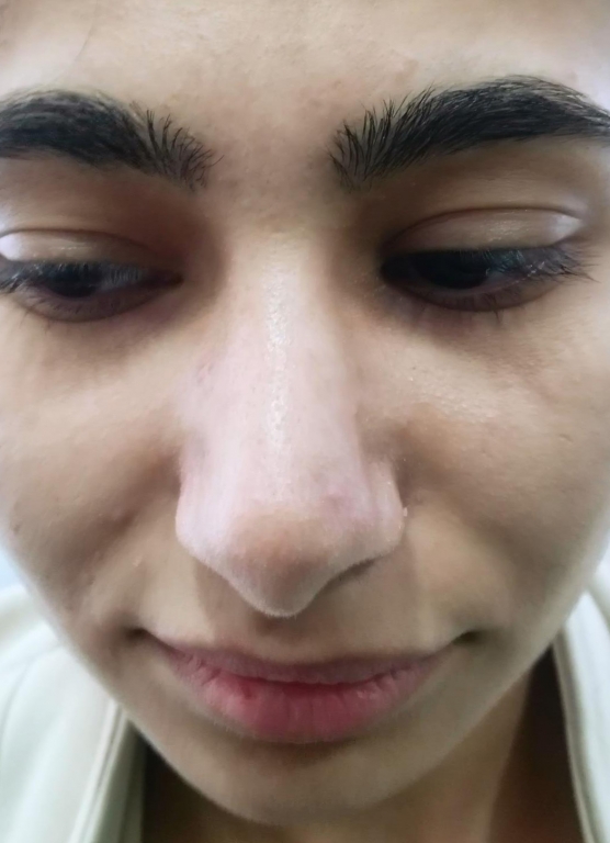
DiscussionNasal muscles innervated by facial nerve is located inside the SMAS layer covering the osteocartilaginous skeleton of the nose. It was noted that during septorhinoplasty operations, this layer may be damaged by open or closed technical septorhinoplasty operations with the dorsal nasal flap. If this layer is damaged, movement of these muscles will be affected, therefore, the nasal movement will be affected and the nose will become paralytic [8]. Synkinetic movements occur between 3-6 months in the process of neural repair after injury [2, 9]. The underlying pathophysiology of synkinesis is very complex and multifactorial. Almost all cases of synkinesis develop as a continuation of nerve trauma [the exception is when it was born as in the Duane-Retraction Syndrome and Marcus Gunn phenomenon]. Synkinesis can be induced by nerve injury, surgical procedures, neuritis, neuroma [10] and physical injury [11]. In rhinoplasty surgery, more attention should be given to the nasal muscles for a more functional and aesthetic result. Operative procedures may partially disrupt the nasal muscular structure [12]. Synkinesis is a recognized complication after peripheral facial nerve palsy. Different types of synkinesis have been described, most commonly oral-ocular and ocular-oral synkinesis. In the literature, ocular-nasal synkinesis after cosmetic rhinoplasty has been reported. Acquired synkinesis usually develops at least 6 weeks after trauma and is believed to be caused by abnormal regeneration of damaged nerves [13, 14]. Özturan et al. [12] suggested that nasal muscle electromyographic activities decreased after rhinoplasty operations with open technique and the surgical procedure had a detrimental effect on the nasal muscle. Kırgezen et al. showed no difference in nasal muscle injury in open and closed rhinoplasty. [15] The basic principle of plastic surgery is respect for tissue and anatomy. Rhinoplasty surgery tends to evolve in generational epochs in recent years. [16] . A complete subperichondrial and subperiosteal dissection technique after rhinoplasty can minimize soft tissue distortion. [17] Synkinesis may be eliminated surgically. Çiloğlu et al.[18] evaluated oculonasal synkinesis in a 20-year-old woman. The patient with congenital oculonasal synkinesis showed that the involuntary muscle function was eliminated by resection of the compressor narium minor muscle [18]. In another study, two female patients who developed synkinesis due to unilateral contraction of medial eyebrow muscles [procerus] after cosmetic rhinoplasty were successfully treated with Botulinum toxin A [1]. In our case, the synkinesis has been eliminated by botulinum toxin A injected in the compressor narium minor muscle [video 1,2,3). Muscle damage should be minimized in rhinoplasty. Performing dissection in the subperichondrial plane can minimize iatrogenic muscle damage and subsequently prevent abnormal reinnervation and synkinesis. Botulinum toxin A injection can effectively prevent unwanted muscle contractions in patients with synkinesis. References
|
|||||||||||
| Keywords : Septorinoplasti , Sinkinezis , Botulinum toksin A | |||||||||||

|