

Authors
|
|||||||||||
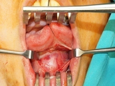
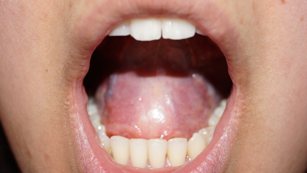
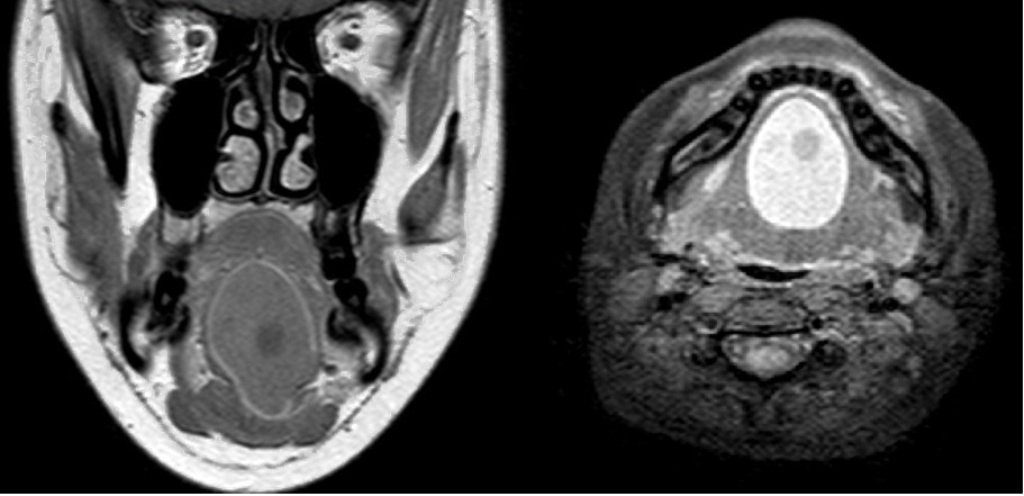
AbstractTeratomas are midline tumors of the pediatric age group. They are mostly located at gonadal or sacrococcygeal region and are diagnosed at young ages. They are rarely seen in the head and neck region. We report a rare case of mature cystic teratoma in the sublingual area and discuss the clinical presentation, diagnosis, and treatment approaches. The 23-year-old female patient presented with a swelling under the tongue that had been present for nearly a year. Radiologic evaluation revealed regular borders, displacing the tongue superiorly semisolid-cystic mass on the floor of the mouth. The mass was totally excised under general anesthesia with an intraoral sublingual mid-line incision. Histopathological examination revealed that the mass was a mature cystic teratoma with hairy sebaceous material in the lumen. In conclusion, we believe that teratoid tumors should also be considered in the differential diagnosis of neck lesions especially if the mass has regular contours and a semisolid-cystic nature radiologically despite the fact that they are.IntroductionTeratomas are midline tumors of the pediatric age group. They are mostly located at gonadal or sacrococcygeal region and are diagnosed at young ages [1,2]. The incidence rate in adults is less than 1%. They are more common in females and two peaks have been reported in the second and third decades [3]. The locations of teratomas incidences in a decreasing order of incidence are the ovaries, the testes, the anterior mediastinum, the retroperitoneum, the presacral and coccygeal regions, the intracranial region and the neck [4]. This article will present the clinical and radiological findings and surgical treatment results of a patient with a localized mature cystic teratoma (MCT) in the sublingual area Case ReportThe 23-year-old female patient presented with a swelling under the tongue that had been present for nearly a year. The physical examination revealed a mass with an approximate size of 4x3 cm located in the sublingual area that was palpable in the submental area and displaced the tongue superiorly and a had regular surface (Figure 1).
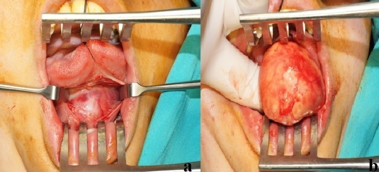
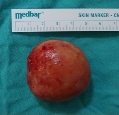
The results of the routine laboratory analysis of the case in our clinic were normal. Surgical excision was planned. The mass was totally excised under general anesthesia with an intraoral sublingual mid-line incision (Figure 3, 4).
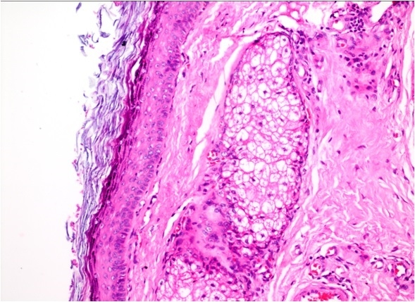
Histopathological examination revealed that the mass was a mature cystic teratoma with hairy sebaceous material in the lumen (Figure 5). No complications developed in the postoperative period or the one-year follow-up period of the patient and no additional treatment was planned.
DiscussionTeratomas are tumors of embryonic origin that comprise components of the three germ layers (endoderm, mesoderm and ectoderm) in various ratios and are rarely located in extragonadal areas. The mediastinum, neck and retroperitoneal areas have been reported in literature as extragonadal locations [2]. In our case, the mass confirmed as a teratoma by pathological analysis was located in a more uncommon area, the sublingual area. Neck teratomas have been rarely reported in literature. Teratomas are childhood tumors and are diagnosed during the early years of life [3,5,6]. The case presented was a 23-year-old female. Cartilaginous, fatty, bony, muscular and neural elements may be grossly visible in histopathological examination [7]. Hairy sebaceous material was observed in the macroscopic examination of the excised material. Teratomas appear as heterogeneous mass with internal calcification in ultrasonography [8]. The ultrasound findings of the case presented are consistent with these findings. The complications of MCT are torsion (16%), rupture (1-2%) and infections. Malign transformation occurs in 0.2-2 % of MCTs and is very rare [9,10]. The golden standard of MCT treatment is surgical excision. It has been reported that the prognosis is favorable in patients treated with total surgical excision [2]. Our case was treated with total surgical excision and no findings suggestive of relapse or late complications were observed during the one-year follow-up. In conclusion, we believe that teratoid tumors should also be considered in the differential diagnosis of neck lesions especially if the mass has regular contours and a semisolid-cystic nature radiologically despite the fact that they are. References
Presented at36th Turkish National Congress of Otorhinolaryngology and Head & Neck Surgery (November 5-9, 2014, Antalya, Turkey). |
|||||||||||
| Keywords : Teratom , Baş boyun tümörleri , Ağız boşluğu | |||||||||||
|