

Authors
Yozgat Şehir Hastanesi
Yozgat Şehir Hastanesi
Yozgat Şehir Hastanesi
|
|||||
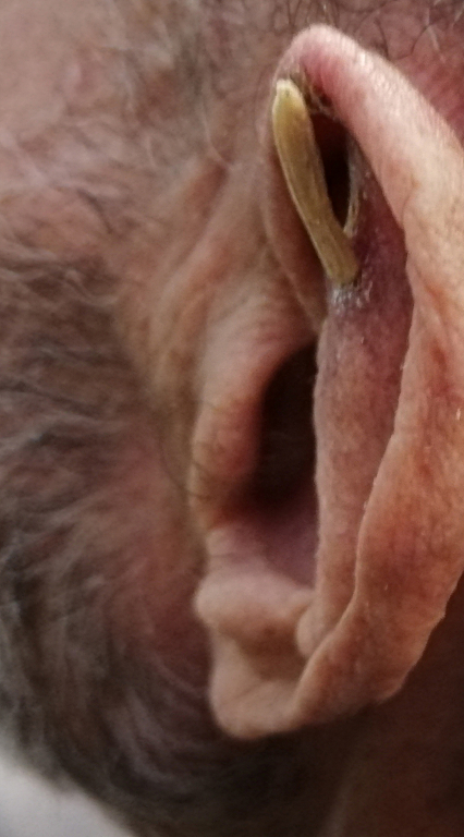
AbstractA case of asymptomatic cutaneous horn occurring in the antihelix part of the left ear was presented in a 77-year-old male patient. Cutaneous horn is a cone-shaped hyperkeratotic lesion. It is more common in sun-exposed areas and older individuals. This case report aims to contribute to the cutaneous horn literature.IntroductionCutaneous horn is a slow-progressing hard keratinized lesion with a horn-like protrusion from the skin. It is a morphological definition rather than a pathological diagnosis. It can also occur as a result of many benign, premalignant or malignant diseases such as seborrheic keratosis, basal cell carcinoma, and squamous cell carcinoma [1]. Histopathologically, there are hyperkeratosis and parakeratosis associated with atypical keratinocytes [2]. Although it can occur anywhere on the body, it is more common in places exposed to the sun. It is most commonly seen on the dorsal surface of the hand and scalp [2]. It was seen as a symbol of witchcraft in modern prehistoric times [3]. In this article, we aimed to review the literature by reporting a case report of cutaneous horn in the antihelix part of the left ear. Case ReportA 77-year-old male patient presented to us with a slow growing hard cutaneous horn in his left ear. On physical examination, there was a hard, immobile and rough surface lesion approximately 2 cm long on the left ear antihelix (Figure 1,2). The patient did not have lymphadenopathy. Cutaneous horn was totally removed from the base by including cartilage tissue with a safe surgical border. In histopathological examination, a lesion involving subcutaneous tissues with hyperkeratotic horn with a size of 2 * 0.5 * 0.3 cm was observed in macroscopy. Microscopically, this lesion has been reported as actinic keratosis. There is scar tissue in the excised area in the 6-month follow-up examination. Complete recovery was achieved without relapse. The patient is still in our follow-up.
DiscussionThe term "cutaneous horn" refers to a slowly progressive keratinized lesion that protrudes a horn-like protrusion from the skin. Usually this protrusion is conical and shaped like a horn. It has been reported in the literature as pointed, grooved and curved shapes [4]. Despite its hard appearance, it does not contain bone tissue. The length of these lesions ranges from a few millimeters to 38 cm [5]. The frequently affected age range is 50-89 [6]. Although it can be seen in different parts of the body, it is most common in areas exposed to the sun. It has also been reported in the literature in areas protected from the sun, such as the penis and groin. The reasons why these lesions occur are still unknown. Radiation, chronic irritation, burn scar, and Human papilloma virus-2 infection can be triggering factors [7,8]. Since cutaneous horn is a morphological definition and can be seen in many underlying diseases (SCC, actinic keratosis, seboraic keratosis, verruca vulgaris, Kaposi's sarcoma etc.), histological diagnosis should be confirmed. For this, a biopsy should be performed from the base of the horn. Histopathologically, it is divided into three types as benign, premalignant and malignant. In the retrospective analysis of 222 cases, Mantese et al. reported that 58.56% of cutaneous horn lesions consisted of malignant or premalignant diseases and 41.44% of them were benign diseases [6]. Gender, age, location and size of the lesion are the main factors associated with morbidity. In this series, the age distribution is between 14-95 years old, and they noted an increase in the prevalence of premalignant lesions after the age of 50 [6]. Yu et al. investigated 643 cutaneous horn cases, 61% reported to be benign and 39% were premalignant or malignant. The most frequently associated premalignant skin lesions are actinic keratosis and Bowen's disease. Yu et al. found that there was higher height-to-base ratio in benign lesions when compared with premalignant or malignant lesions, suggesting that a cutaneous horn with a wider base is more likely to be associated with malignancy or premalignant lesions [9]. It takes a long time for the development of the disease, and the larger the base of the lesion, the higher the risk of being premalignant or malignant [10].In the analysis of Yu et al, it was found that a cutaneous horn on the face is 2 times more likely to be premalignant or malignant than other parts of the body [9]. The clinical preoperative diagnosis of cutaneous horn is suspected based on the antler-like appearance of the tumor apex, but assessment of the underlying potentially malignant base is clinically important [10]. There are no reliable parameters to clinically predict malignancy. Therefore, in cases of cutaneous horn, completely wide resection of the tumor base is required. It should be remembered that cutaneous horns can be seen with benign, premalignant or malignant lesions. Malignancy should be kept in mind especially in fast developing lesions and those with a large base. Surgical excision should be the first choice in treatment and histopathological examination should be performed. Definitive diagnosis can only be made after pathological evaluation. AcknowledgementOp. Dr. Erol Erkan'a yardımlarından dolayı teşekkür ederiz References
|
|||||
| Keywords : Cutaneous , Horn , Antihelix | |||||


|