

|
|||
AbstractAdenotonsillar disease is one of the most common diseases of childhood. Children suffer from upper airway obstruction due to adenotonsillar hypertrophy as well as attacks of infection. Its clinical presentation ranges from mild recurrent infection to congestive heart failure. Williams-Beuren Syndrome (WS;OMIM 194050), also called Williams Syndrome is characterized by dysmorphic facial features, neurological findings and endocrinological abnormalities resulting from various lengths of deletions in the q11.23 region of the 7th chromosome. We present unproblematic adenotonsillectomy under general anesthesia in a child with WS and adenotonsillar hypertrophy.IntroductionAdenotonsillar hypertrophy (ATH) is a common cause of upper airway infection, upper airway obstruction, and obstructive sleep apnea (OSA) in children. Patients with WS characteristically have dysmorphic facial features, neurological findings, endocrinological abnormalities also heightened musical and verbal abilities, but low visuospatial abilities and IQ. Cardiovascular anomalies are the most common manifestations of WS and CVS disorders are found in 60-85% of patients with WS. The majority of sudden-death cases have been associated with myocardial infarction [1] The risk of sudden death from cardiomyopathy is 25–100 times greater than the normal population. Systemic hypertension (55–60%of patients with WS) has been found to be associated with hypercalcemia (15% of patients) and narrowing in renal vessel (60% of patients) due to defect in elastin protein [2] The reduced elastin production leads to noncompliant vessels, which reduces diastolic aortic recoil, which inturn, lessens coronary blood flow. Also arterial walls are exposed to multilayer thickening, especially in high-pressure arteries. These subsequently lead to upcoming intimal lesions causing stenosis or occlusion.
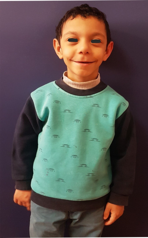
We hereby present the results of adenotonsillectomy and bilateral paracentesis in a patient with WS who presented with chronic serous otitis media and severe airway obstructive symptoms. We wanted to draw attention to the importance and affects of upper airway obstruction and other comorbidities on general anesthesia in WS patients. According to our literature knowledge, it is the first reported uneventful adenotonsillectomy case performed under general anesthesia in WS patient. Case ReportOur patient was a 7-year-old boy with advanced upper respiratory problems. The diagnosis of WS had been made via fluorescent in situ hybridization (FISH) which showed signals indicating single-allele deletions in the 7q11.23 region. The patient had undergone abdominal ultrasound evaluation in 2014 which showed no significant problems. In 2019, echocardiography had showed the presence of mild left ventricular hypertrophy, supravalvular aortic stenosis, and mild mitral regurgitation. The patient’s general physical examination revealed significant growth retardation.(Figure 1)
When a detailed medical history was taken, we found that the patient had continuously applied to various centers with upper respiratory complaints; although surgical intervention was planned, surgeries were delayed or not scheduled due to significant risks associated with Williams syndrome. The patient applied to our center with similar upper respiratory complaints. In addition to significant growth retardation, the patient was found to have bilateral tonsillar hypertrophy, chronic rhinosinusitis and bilateral otitis media with effusion at the initial physical examination. Chronic rhinosinusitis therapy was initiated with a 1-month antibiotherapy and a follow-up visit was scheduled. In the control evaluation, it was determined that the patient’s obstructive symptoms and chronic rhinosinusitis had regressed and the patient’s family confirmed that obstructive problems had become less of a problem after the treatment. At the end of this follow-up visit, the patient’s family was informed that surgical intervention would be beneficial for these chronic problems and the risks of such surgery in patients with WS was thoroughly explained. After the family stated that they intended to follow this surgical path of treatment, surgery was scheduled. The informed consent was obtained from the mother and father of the patient to perform this procedure. Patient's preoperative blood pressure 100/50 mmHg, heart rate 100 p/min, body temperature 36.5 °C, oxygen saturation with pulse oximeter 98%. Induction of anesthesia has been started with low dose propofol and rocuronium bromide and maintenance balanced anesthesia has been proved by intravenous propofol infusion. Inhalation anesthesia was not used. Patient has been monitorized during the operation and blood pressure, pulse, oxygen and carbon dioxide saturations and body temperature were checked frequently. Cardiac monitoring for ST segment changes were followed for risk of myocardial infarction. Patients has been reanimated just after the end of surgical operation. Vital signs were stable postoperatively. Postoperative blood pressure 110/70 mmHg, heart rate 96 p/min, body temperature 36.6 °C, oxygen saturation with pulse oximeter 97. Adenoidectomy under general anesthesia was performed using 'cold knife technique' dissection. Then, bilateral tonsillectomy and hemostasis were performed using bipolar cautery with an estimated blood loss of 10 ml. Finally, bilateral paracentesis to tymphanic membranes was performed. The vital signs were stable throughout the procedure. The operation was completed in 50 minutes. Dissection samples were sent for histopathological analysis. In the current case, the patient’s surgery was scheduled after careful consideration with the cardiology and anesthesiology departments after informed consent of the patient’s family. His surgery was successful and no complications were observed during surgery and follow-up. The vital signs were stable. He was discharged the following day. The postoperative course was uneventful and in third week appointment, he was found to be symptom-free. DiscussionAccording to our literature knowledge, this case is the first reported uneventful adenotonsillectomy case performed under general anesthesia in WS patient. Respiratory acidosis and pulmonary vasoconstriction due to hypoxemia and hypercarbia may be observed in patients with adenotonsillar hypertrophy due to chronic upper airway obstruction. Therefore, WS patients with adenotonsillar disease should be followed closely from an early age due to cardiac anomalies. Adenotonsillectomy is the primary treatment for pediatric OSAS with adenotonsillar hypertrophy. In our case, our patient was seven years old and he had supravalvular aortic stenosis accompanying mild left ventricular hypertrophy and mild mitral regurgitation. Congenital heart diseases are found in 60-85% of patients with WS. An optimized anesthesia protocol has not been established for every patient with WS. Clinical evaluations of the patients and the type of procedure are the main factors that will shape the anesthesia protocol and ideal sedation level. During anesthesia induction, neuromuscular blockers are used combined with general anesthesia to facilitate tracheal intubation and relaxation of skeletal muscles. We used low dose propofol and rocuronium bromide for induction of anesthesia and maintained the balanced anesthesia with intravenous propofol infusion. Due to the profound vasodilator effect of propofol, its careful use has been recommended in WS patients. Propofol exerts negative chronotropic and negative inotropic effects on human cardiac muscle cells in a dose-dependent manner [3] There was no intubation difficulty in our case. WS patients are under 3 years of age with biventricular outflow tract obstruction (BVOTO), coronary artery involvement, carry the highest risk of cardiac complications for general anesthesia and sedation procedures. In addition, it has been reported that SVAS poses a great risk for myocardial ischemia when diastolic blood pressure falls further decreases coronary blood flow [4]
Our case was over 3 years old and had an isolated SVAS anomaly. In addition to adenotonsillar disease, bilateral serous otitis media was present. We have assumed that age related risk was lower for our patient and only an isolated supravalvular aortic stenosis helped our patient to better tolarate the given anesthetics . Therefore while administering an anesthetic, goals must be establishing a balance between myocardial oxygen supply and consumption, maintaining the age appropriate heart rate, sinus rhythm, myocardial contractility, ideal systemic vascular resistance (SVR), avoiding increases in pulmonary vascular resistance. Also anesthetics minimize the reduction of diastolic blood pressure and maintain coronary blood flow, adequate hydration to augment leftventricular filling pressures are other key factors for the anesthetic management of WS patients with cardiovascular abnormalities. Tachycardia, hypertension, and hypotension should be prevented during the perioperative stage. Suggestions for WS patients are similar to those for adults with ischemic heart disease. An ECG should be obtained perioperative period and importantly right before any anesthetic intervention to determine heart rate and rhythm, electrical conduction and repolarization (QTc) and the presence of any ischemia [6] A prolonged corrected QT (QTc) has been reported in 14% of patients with WS. This is thought to be caused by myocardial ischemia. The prolongation of the Q-T interval in patients with Williams syndrome is similar to the increase in the Q-T interval in patients with ischemic heart disease [7] We did not observe any increase in the Q-T interval in our case. For inhalation inductions, should be aware of the risk of deep peripheral vasodilation at the high concentrations necessary to induce anesthesia adequately. Failure to administer vasoactive agents or IV fluids during induction increases the risk of hypotension and cardiovascular collapse may occur if rapid IV access is not achieved [8] Barbiturates (thiopental, pentobarbital) has ability to reduce SVR and other agents reduce SVR and myocardial depression are inhalation anesthetics, profolol, and benzodiazepines (midazolam, lorazepam).Even low and gradual increases in sevoflurane concentrations have been reported to cause cardiac arrest in high-risk patients [4] Ketamine increases heart rate and myocardial oxygen consumption while preserving myocardial contraction and SVR. Etomidate has also been used successfully in high risk patients. Patients may show a variable response to neuromuscular blockade. The use of the depolarizing agent succinylcholine is somewhat controversial because of concern for the exaggerated hyperkalaemic response seen in other myopathic conditions [9] Adenotonsillectomy patients are still at high risk for airway obstruction, laryngospasm, desaturation, pulmonary edema and respiratory arrest subsequent to extubation, even when patients are extubated in an "awake" state Conclusion In conclusion, adenotonsillectomy in pediatric WS patients >3 years of age, may result in an uneventful intraoperative and postoperative outcome with an appropriate general anesthesia protocol, as in our case. When adenotonsillar patients with WS come to the otorhinolaryngology clinic, pre-anesthesia evaluations in terms of pediatric cardiology, pediatric endocrinology and anesthesia should be made meticulously and risk factors should be determined. In WS patients with adenotonsillar hypertrophy, the operation should be planned after 3 years of age due to high risk of complication. References
|
|||
| Keywords : Williams sendromu; Risk; Genel anestezi ; Adenoidektomi; Tonsillektomi | |||

|