|
|||||
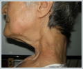
AbstractSwallowing is a complex, multiphase event and various disorders can cause pharyngeal dysphagia particularly at old age. Hence, diagnosis and management of pharyngeal dysphagia in geriatric patients require detailed analysis and multidiciplinary management. We present a unique case of multifactorial pharyngeal dysphagia due to a cricopharyngeal bar and a severe cervical kyphosis in a 66-year old woman.IntroductionSwallowing is a complex, multiphase event and various disorders can cause pharyngeal dysphagia. Cricopharyngeal (CP) muscle abnormalities have been implicated as significant causes of dysphagia in the elderly population [1,2]. CP bar is a radiological term defining a posterior indentation or a persistent prominence of the CP region at the level of the cricoid cartilage that narrows the PES during a barium swallow [1]. It is one of the potential etiologies of pharyngeal dysphagia that is frequently encountered by otolaryngologists. Open CP myotomy has long been accepted as a treatment option for patients with CP bar, however, more recently, endoscopic laser-assisted CP myotomy (ELCPM) has become the technique of choice for many head and neck surgeons. Since swallowing is a complex event, various disorders, alone or combined, may lead to dysphagia. It is very well known that dysphagia can be associated with postural disorders, craniovertebral abnormalities or spine surgery. Disorders of spine such as scoliosis, hyperlordosis, kyphosis, anterior cervical osteophytes have known to usually cause mechanical obstruction by compressing the aerodigestive lumen, and thus, impair the passage of bolus [3,4]. We present an interesting and unique case of multifactorial pharyngeal dysphagia and comment on the potential challenges in the diagnosis and management of pharyngeal dysphagia in the elderly population. Case ReportA 66-year old woman presented to our department for ELCPM for CP bar (Figure 1).
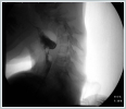
We agreed that her dysphagia was due to kyphosis and CP bar which was diagnosed by videofloroscopy (Figure 2).
The patient was informed that an ELCPM followed by swallowing therapy may help at some degree but, kyphosis should be corrected in order to reach best swallowing outcome. Consequently, she was referred to our spine surgery team for surgical correction of her kyphosis. After discussing the benefits and risks of surgery with spine surgeons, patient preferred to try CP myotomy first and be followed by our swallowing team with conservative treatment including swallowing therapy and puree-liquid diet. The risks and benefits of this surgery was discussed in detail and an ELCPM was performed. A hard CP bar and a dilated/enlarged pharynx above the bar were noted. We noticed a more fibrotic/scarred CP region than usual that may be attributed to previous irradiation and dilatation. Postoperative course was uneventful and she tolerated soft diet two days after surgery. She was discharged that day and instructed to modify her diet gradually from soft to regular diet within a couple weeks. Unfortunately, she came back 6 weeks after surgery with difficulty swallowing just like before the CP myotomy. DiscussionDiagnosis and management of pharyngeal dysphagia in geriatric patients can be challenging for all clinicans dealing with swallowing disorders. Our patient had a CP bar obstructing the bolus passage, however, the main problem might have been that she was not able to generate enough pharyngeal pressure to push the bolus down because the cervical spine was too far away from the base of tongue and the pharynx was too large to squeeze the bolus. It would be better if this was confirmed by pharyngeal manometry that was not available. Even though we had seen that the impact of kyphosis was much more overwhelming, we had expected that myotomy would help her to tolerate her soft diet but, it did not work as much as we expected. Laurikainen et al. [5] have investigated muscle pathology in idiopathic CP dysphagia in a small cohort of patients and found different immunohistochemistrical and electron microscopical changes in each of their patients. Inspite of the great variety of possible etiologies causing idiopathic CP dysphagia in their patients, they noticed both subjective and objective benefit from CP myotomy in all of their patients. Maybe, the success of our myotomy was minimized by the CP scar secondary to previous irradiation and dilatation. Kyphosis is frequently seen in the elderly population but dysphagia due to kyphosis is extremely rare. In a pubmed search, we were able to find two case reports. In the first case, dysphagia was caused by esophagial atony due to cervico thoracic kyphosis, [6] whereas kyphosis resulted in a 90-degree bend in the esophagus leading to dysphagia in the second one [7]. In our patient, interesting enough, isolated cervical kyphosis was not obstructing the lumen, it was even enlarging the pharyngeal space in the sagital plane. Excessive enlargement and strain in the pharyngeal lumen was why she had difficulty swallowing. Otolaryngologists and those who are dealing with pharyngeal dysphagia should keep in mind that swallowing is a multiphase event and various disorders at a time can cause pharyngeal dysphagia. Not only the obstruction/narrowing of the pharynx but also the strain/excessive enlargement of it can cause pharyngeal dysphagia. Therefore, pharyngeal dysphagia patients require detailed analysis and multidiciplinary management. We believe this report will remind clinicians that dysphagia can be multifactorial, particularly at old age, and the impact of the angle of cervical spine on pharyngeal space should be meticulously inspected in pharyngeal dysphagia patients in addition to all other swallowing studies. References
Presented atThis case has been presented as e-poster in ‘15. Türk Rinoloji Kongresi ve 7. Ulusal Otoloji Nörootoloji ve 3. Ulusal Baş Boyun Cerrahisi Kongresi’, Antalya, Turkey (April 4-7, 2019). |
|||||
| Keywords : Disfaji , geriatri , krikofarengeal bar , kifoz , servical kifoz | |||||
|


