

Authors
|
|||||
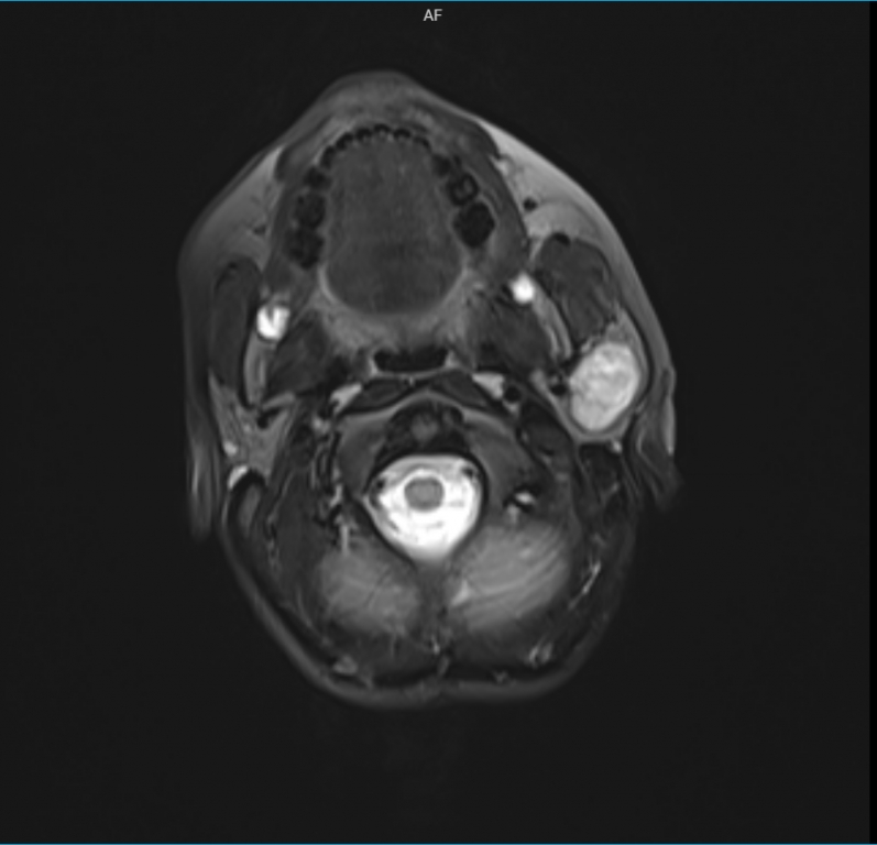
AbstractTumors of the salivary glands are not that rare and constitute an important field in oral-maxillofacial pathology. The diversity of histological aspects displayed by neoplasms of the salivary glands causes great difficulty in terms of universal classification. They are more common in women than in men and are more common between the ages of 30-50. Less than 5% of the affected population is pediatric. In this study, a case of an 8-year-old patient with pleomorphic adenoma arising from the deep lobe of parotid with perineural invasion is presented in the light of current literature.IntroductionPleomorphic adenoma is the most common epithelial tumor of the major salivary glands. 65% of all salivary gland tumors are pleomorphic adenomas [1]. Pleomorphic adenoma originating from large salivary glands is mostly in parotid (90%) gland, rarely in submandibular salivary glands; those arising from small salivary glands are mostly localized in the palate. They are more common in women than in men and are more common between the ages of 30-50 [2]. Salivary gland carcinomas are exceedingly rare in the pediatric population. No other case of pleomorphic adenoma arising from the deep lobe of parotid with a perineural invasion of a pediatric patient was reported in the literature. In this case report, an 8-year-old child with pleomorphic adenoma arising from the deep lobe of parotid with perineural invasion is presented in the light of current literature. Case ReportAn eight-year-old female patient approached our clinic with swelling under her left ear that had been present for the past one year. The swelling had gradually increased to its present size and was erythematous. The swelling was firm, non-tender, and affixed to the surrounding structures. The facial nerve examination was normal. Magnetic resolution imaging (MRI) and computerized tomography (CT) of the lesion were requested both to evaluate bony structures and soft tissues better. MRI T2-weighted images exhibited a 25 mm × 15 mm sized, well-defined, high-intensity, heterogeneous mass arising from the deep lobe of the left parotid, which had displaced the surrounding soft tissue (Figure 1), and CT showed a 27 mm × 22 mm sized mass arising from the deep lobe of the left parotid.CT showed no bony destruction.
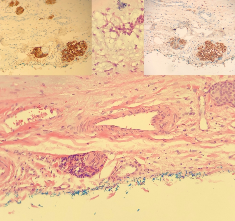
Fine needle aspiration cytology (FNAC) was consistent with pleomorphic adenoma. Routine blood results were within the normal range. The patient subsequently underwent total parotidectomy with preservation of the facial nerve under general anesthesia. The postoperative pathology result was compatible with pleomorphic adenoma. But some foci had perineural invasion and vascular invasion. (Figure 2). The patient was followed up closely without radiotherapy or additional surgery.No recurrence has been observed in one year of follow-up.
DiscussionPleomorphic adenoma is a mixed tumor originating from epithelial and mesodermal elements of salivary glands. [3]. The majority of these tumors that frequently develop in the parotid gland originate from the superficial lobe and are in the form of a symptom-free mass lesion. In our case, the tumor arose from the deep lobe. Most cases with parotid tumors present with a history of a slowly enlarging mass, as our patient, or are incidentally discovered by physicians during a routine examination [4]. Some reports suggest ultrasound imaging as an initial investigation to differentiate cystic/vascular lesions from solid masses. However, MRI is a more comprehensive diagnostic tool that helps in preoperative assessment of the local-regional extent of the mass, detection of the facial nerve and regional lymph node involvement, and differentiation from other malignancies/vascular tumors [5]. CT could be used to evaluate tumor extension and invasion. In addition, CT and MRI are helpful in differentiating between deep lobe tumors and parapharyngeal tumors [6]. Ultrasound-guided FNAC helps in determining the subtype of the parotid tumor based on histopathology. It is crucial that CT should be applied as the last option for children because of radioactivity. In our case, both MRI and CT were applied to plan the surgery in detail, but in fact, surgery could have been done without CT imaging. Pleomorphic adenoma is the second most common neoplasm of the parotid gland after hemangioma, representing 40% of all benign parotid tumors. But as far as we searched the literature, in the pediatric group there is no case of pleomorphic adenoma arising from the deep lobe of the parotid and causing perineural invasion reported. “Invasion” of peripheral nerves by epithelial cells used to be regarded as a feature of malignancy. However, an analogous phenomenon was subsequently described in benign lesions of the breast, prostate, pancreas, gallbladder, endometrium, skin, and salivary glands. Histologically, pleomorphic adenoma contains epithelial, myoepithelial cells, mesenchymal or stromal elements. It is divided into histologically different types according to the cellularity and stroma rates in the tumor [7]. It constitutes 70-85% of all parotid tumors. Malignant masses are extremely rare. Simple enucleation is not accepted as the right approach because it carries a risk of recurrence between 30-50%. For this reason, the most limited procedure that can be surgically performed in the pleomorphic adenoma in the parotid should be a superficial parotidectomy but we performed a total parotidectomy in our case because of a tumor arising from the deep lobe. Also, adjuvant radiotherapy is considered in cases of incomplete surgical resection, persistent lymph node involvement, perineural invasion, or an aggressive histological grade tumor. However, the risk of post-irradiation complications such as facial/dental deformities, secondary malignancies, trismus, and hyposialia should be carefully evaluated [8]. In our case, the perineural invasion was reported as a pathological diagnose, and the patient was kept under close follow-up without radiotherapy considering the side effects and patient's age, and there was no recurrence in the 1-year control. In conclusion, our case report is unique due to the absence of a report of pleomorphic adenoma arising from the deep lobe of the parotid and perineural invasion coexistence in childhood. Also, without an additional treatment considering the age group, we observed no recurrence till the first year of surgery. Informed ConsentInformed consent was obtained from the individual participant's family members included in the study before surgery.References
|
|||||
| Keywords : Neoplazm , Pleomorfik adenom , Çocukluk çağı , Perinöral invazyon | |||||

|