

Authors
|
|||||
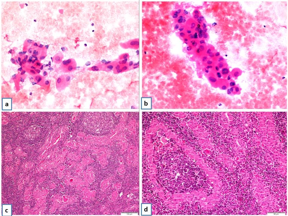
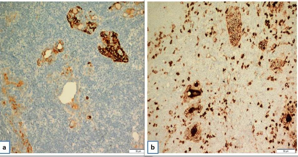
AbstractWarthin-like papillary carcinoma of the thyroid is a recently described rare variant of thyroid papillary carcinoma. Although it is commonly associated with lymphocytic thyroiditis, the histopathologic features resemble the Warthin tumor of the salivary gland. Therefore, the final diagnosis is confirmed by histopathological examination. The prognosis is similar to conventional papillary carcinoma. We present a case of Warthin-like papillary thyroid carcinoma in a 29-years-old woman.IntroductionWarthin-like papillary carcinoma of the thyroid is a recently described rare variant of thyroid papillary carcinoma. It's most prominent histologic property is its papillary structure, formed by neoplastic cells, which have lenfoid stroma, and oncocytic papillary carcinomatous nuclear properties [1]. This tumour was first defined by Apel and colleagues in 1995, and named after its morphologic appearance resembling Warthin tumour of the salivary gland [2]. ITs prognosis is similar to classical papillary carcinoma [3-6]. We present a case of Warthin-like papillary thyroid carcinoma in a 29-year-old female patient and describe the clinicopathological features along with the differential diagnosis of this rare variant. Case ReportA 29 years old female patient presented to the general surgery clinic with a neck mass, growing over the last year. Ultrasonography (USG) of the neck revealed a large thyroid gland, a 20x15 mm large hyperechoic nodule in the left lobe with complete peripheral halo and significant vascularization. There were no enlarged lymph nodes. There was no previous exposure to radiation. Thyroid function test results were normal. However, anti-TPO antibody levels were found to be increased (312.0 UI/ml, range: 0.0-10.0 UI/ml). These findings were consistent with chronic otoimmune thyroiditis. A fine needle aspiration of the nodule showed papillary structures lined by wide cells with granular cytoplasm, which form irregular clusters within a lymphocyte-rich stroma with cytologic findings of classical papillary carcinoma (Figure 1a-1b). Due to cytologic malignant characteristics, total thyroidectomy was performed. Macroscopic examination of the total thyroidectomy material showed a 20x15 mm solid, white-greyish mass with regular contours, located at the left lobe, confined to the thyroid capsule. Microscopic examination showed a tumour formed by papillary and follicular structures, including neoplastic cells with prominent germinal centers outlined by lenfoid stroma, abundant granular eosinophillic cytoplasm, pseudoinclusions and nuclear grooves (Figure 1c-1d). Immunohistochemical examination showed positive staining with cytokeratin 19 (CK19) and Hector Battifora Mesothelial Cell (HBME-1) (Figure 2a-2b).The surrounding thyroid tissue showed features of lymphocytic thyroiditis, and a 2 mm size follicular variant of papillary microcarcinoma. There was no lymphatic vascularization, and the final diagnosis was Warthin-like papillary thyroid carcinoma. The patient received radioactive iodine therapy. At one year follow up, the patient was disease free and thyroglobulin levels were undetectable.
DiscussionWarthin-like papillary carcinoma is rarely encountered variant of papillary carcinoma of the thyroid [1]. Compared to classical papillary carcinomas, they appear at younger ages and in women. Among the 54 patients reviewed in the current literature, the mean age was 50 years and there were only five males. Its clinical presentation is not different than the other differentiated thyroid tumours. If the tumour is small, signs and symptoms may be subtle. If the tumour enlarges, thyroid gland may become palpable, and dysphagia may occur. Clinical signs and thyrid hormone levels depend on the presence and severity of thyroiditis. Also, USG and computerized tomographic (CT) findings are not different than the classical papillary carcinoma [7]. Fine needle aspiration may aid in the diagnosis of Warthin-like papillary carcinoma [8]. Cytologic features may show follicular cell clusters and papillary fragments with fibrovascular cores, and a predominance of lymphocytes and plasma cells a with bloody background. Follicular cells may show characteristic features of classical papillary carcinoma (nuclear chromatin clearing, grooves, membrane thickening, and intranuclear pseudoinclusions) and oncocytes (round nuclei with coarse chromatin and prominent nucleoli). All these findings suggested a diagnosis of classical papillary carcinoma, Hashimoto's thyroiditis, or both [7]. Intranuclear inclusions are known to be scarce in Hashimoto's thyroiditis, yet sometimes pseudoinclusions may appear as formalin fixation artefacts, too [8]. As for cellular atypia, oncocytic changes, and nuclear grooves, they may be observed in the absence of follicular cells with pseudoinclusions, and in some other benign cases [9]. All of these factors should be kept in mind to prevent a mistaken diagnosis. It should not be forgotten that Warthin-like papillary carcinoma may also resemble follicular adenoma or carcinoma with oncocytic changes, in the absence of ground-glass appearance and papillary structures [8]. Histological appearance of Warthin-like papillary carcinoma may resemble other variants of papillary carcinoma, such as tall cell or columnar cell variants, both of which are more aggressive compared to papillary carcinoma. Medullary carcinoma, too, may show oncocytic changes. Hurthle cell carcinoma of the thyroid has a worse prognosis compared to papillary carcinoma, and shows prominent oncocytic changes, while it lacks nuclear characteristics of the papillary carcinoma. Warthin-like papillary carcinoma may be differentiated from all these variants by the presence of papillary stalks within a lymphocytic stroma [1]. Just like the classical papillary carcinoma, both of these variants are positive for CK19, HBME-1, Galectin-3, TTF-1, and thyroglobulin [7]. The specimen obtained from our patient was positive for CK19 and HBME-1. However, since the role of immunohistochemical methods is limited in the diagnosis of Hurthle cell and tall cell thyroid cancers, an obvious morphological pattern may suffice for the diagnosis. Based on the current literature, prognosis of Warthin-like papillary carcinoma is similar to the classical variant papillary carcinoma. All cases resolved with cure, however, few reports included patient follow up longer than 3 years. This variant has a low rate of lymph node metastasis. Current literature reports lymph node metastasis in only 12 out of 54 cases (22%), and a lower rate of lymph node metastasis compared to the classical variant [4, 7]. If the metastatic lymph node is closely associated with the thyroid gland, it may be difficult to diagnose the metastasis due to the lymphocytic stroma [10]. Only two cases with focal anaplastic changes were reported, and both of them showed good prognosis [11, 12]. Treatment of Warthin-like papillary carcinoma is similar to the classical variant papillary carcinoma. The absolute diagnosis of this tumour requires histopathological examination of the thyroidectomy material. Treatment includes raidoactive iodine therapy after resection of the tumor. Conclusion Warthin-like papillary carcinoma is a recently defined, rare variant of thyroid papillary carcinoma, and is frequently associated with lymphocytic thyroiditis. Fine needle aspiration may aid in the preoperative diagnosis. Due to the presence of oncocytes and lymphocytic infiltration, it possesses diagnostic difficulties to distinguish from lesions of thyroid, both benign and malignant The absolute diagnosis requires histopathological examination of the total thyroidectomy material. Although its prognosis is very good, long term patient follow up data are needed to have a better understanding of its biological behaviour. References
|
|||||
| Keywords : Warthin tümörü , Papiller karsinom , Tiroid | |||||

|