

Authors
|
|||||||
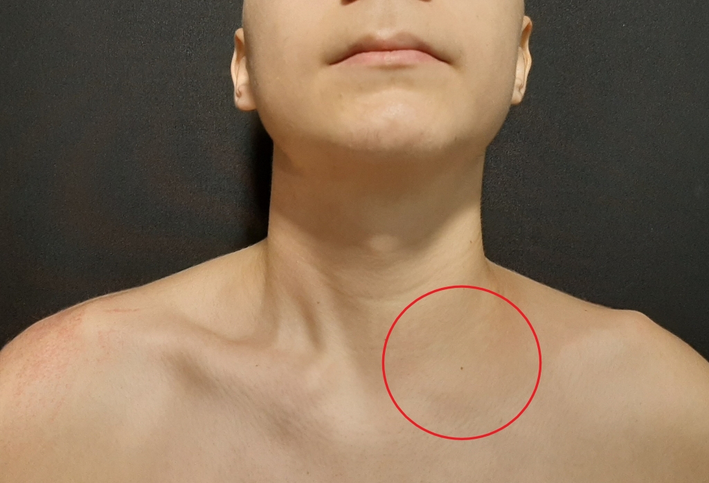
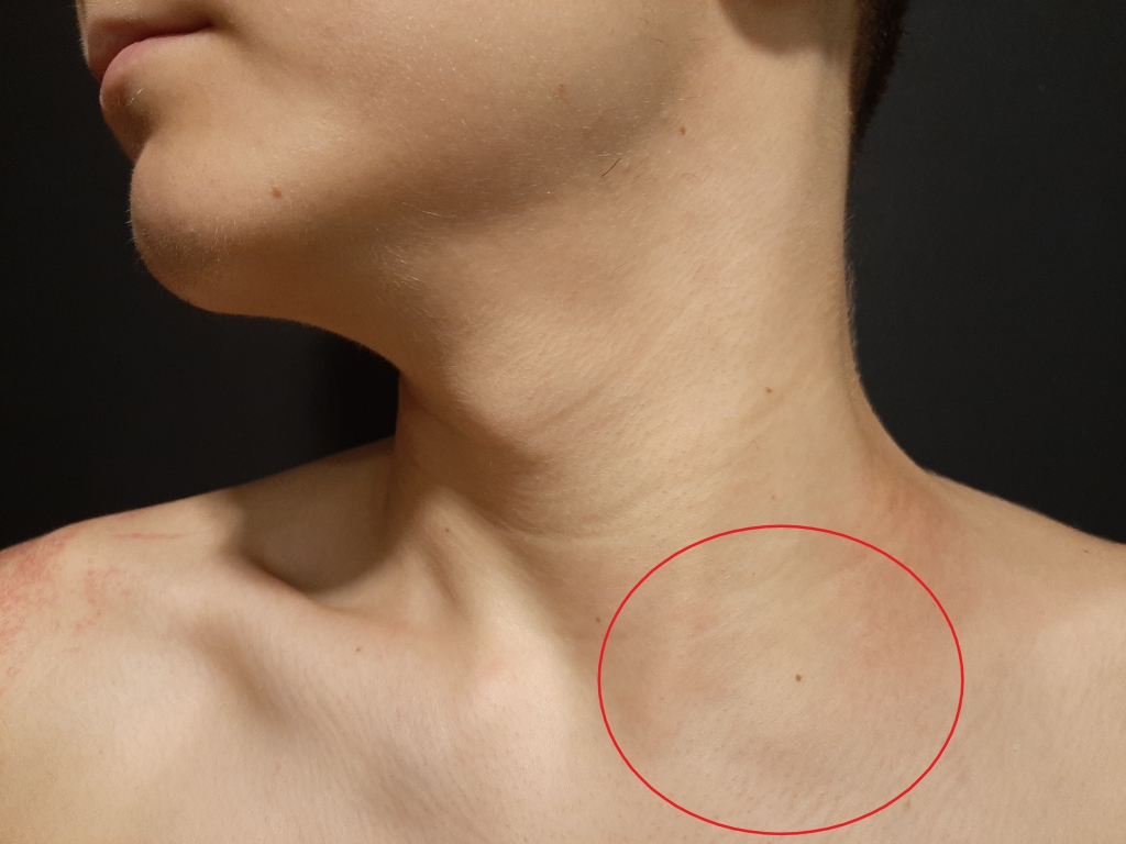
AbstractSternocleidomastoid (SCM) muscle hematomas are rare benign disorders manifested as painy neck swelling. Trauma history, surgery, comorbid illnesses, drug usage, and bleeding disorders must be questioned in the anamnesis. Ultrasonography can be helpful in differantial diagnosis. The general treatment approach is antibiotherapy and follow-up. Surgical management is an option only if there is airway compromise, compression of neurovascular structures or if there is infection.In our case, factor 7 deficiency was detected in an 18-year-old male patient with spontaneous isolated sternocleidomastoid muscle hematoma. Our diagnosis and treatment approach is presented with the current literature. IntroductionAdult neck masses can be due to numerous of disorders, varying between benign lesions to lifethreatining abcess, metastases. Although most common disorders are benign lymphatic lesions or congenital cysts, lots of rare disorders can be manifest as neck masses. After anemnesis and physical examination; usually radiological imaging, labarotuary test and, if indicated, pathological evaluation needed. Neck haematomas are generally associated with trauma, bleeding diathesis, invasive procedure or surgery. They commonly occurred in the anterior triangle of the neck[1]. Spontaneus isolated sternocleidomastoid muscle hematoma is a rare entity and has been described in the literature in only a few cases. In this described cases , hematoma has been observed during thrombolytic therapy or in patients taking aspirin [2,3]. In our case, factor 7 deficiency was detected in an 18-year-old male patient with spontaneous isolated sternocleidomastoid muscle hematoma. Our diagnosis and treatment approach is presented with the current literature. An informed consent was obtained from the patient. Case ReportAn 18-year-old male patient was admitted to our emergency department with complaints of swelling and pain on the left side of the neck. His complaints were present for a week and antibiotherapy (ampicillin-sulbactam 2*1 gram, intramuscular) was started in another hospital with diagnosis of lyhmpadenitis. His complaints did not regress despite antibiotherapy and he has had difficulty in neck movements for last two days. There was no history of trauma, chronic disease or drug use. Also he was not smoker. The patient had a hard, non-fluctuating, ~ 5 cm diameter painful swelling in the left supraclavicular region (Figure 1A,Figure 1B). All ear nose and throat examination, nasopharyngeal and laryngeal endoscopic examinations was normal.
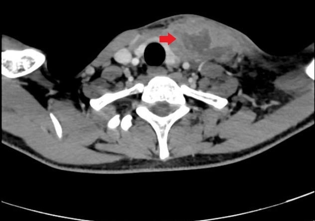
He has an elevated white blood cell (WBC) count of 11,000/uL (normal range: 3.5-11.0 × 10ˆ3/uL) as well as an elevated C-reactive protein of 162 mg/L (normal < = 8.0 mg/L). Coagulation profile revealed prothrombin time (PT) was 19 seconds (normal range: 11-15 seconds) , international normalized ratio (INR) was 1.46 (normal range: 0.8-1.2) , with normal activated partial thromboplastin time(aPTT). Neck ultrasonography showed a loculated fluid collection in the lower third of the left sternocleidomastoid muscle. As the clinic appearance was not typical in terms of abscess, contrast-enhanced neck tomography was used. In the left supraclavicular region, a peripheral contrast-enhanced central hypodense loculated collecting area of 83x22 mm was observed (Figure 2). Approximately 10 cc hemopurulent fluid was drained with ultrasound guided drainage. Ceftriaxone (2*1 g) and clindamycin (3*600 mg) therapies were started. Drained material culture result was negative. Hematology consultation was performed due to prolonged PT and high INR value . With the recommendation of hematology department Factor 7 levels were investigated and detected as low (31%). So the patient was diagnosed with Factor 7 deficiency. Antibiotherapy was completed for 14 days. Clinic findings completely recovered and no additional surgical intervention required.
DiscussionAdult neck masses are frequenlty seen disorders in otorhinolaryngology daily practice. Although most commonly seen disorders are benign in nature, probability of malign etiology increases importance of evaluation. We discussed a sternocleidomastoid muscular hematoma case, manifested by a neck swelling, due to factor 7 deficiency. In reported cases, neck haematoma was associated with trauma, bleeding diathesis, invasive procedure or surgery. Spontaneous neck hematomas are seen in patients with anticoagulant use and most commonly in laryngeal, retropharyngeal and sublingual regions [1]. In the literature, spontaneous isolated sternocleidomastoid muscle hematoma cases are associated with predisposing causes such as thrombolytic therapy and aspirin use. Spontaneous regression was observed all [2,3]. Additionally, sternocleidomastoid muscle hematoma due to weight lifting exercise was treated succesfully with antibiyotherapy without surgicall drainage has been reported [4]. The spontaneous hematomas usually resolve in 2–4 weeks by natural absorption. Surgical management is an option only if there is airway compromise, compression of neurovascular structures or if there is infection [1]. In our case, we performed needle drainage in order to exclude abscess formation and take culture. Surgical intervention was not performed due to absence of airway compression findings and early response to drainage and antibiotherapy. In our case, Factor 7 deficiency was detected. Factor 7 is a protein located in the extrinsic pathway in the coagulation mechanism. Factor 7 deficiency is a rare hereditary coagulation disorder with a prevalence of 1/300.000-500.000. Prolonged prothrombin time (PT) and low Factor7 levels are important laboratory findings for diagnosis. Activated prothrombin time (APTT) value is usually normal [5]. Factor 7 deficiency is usually characterized by an Factor 7 level below 70% (0.7 IU/mL),but clinically relevant manifestations mainly appear when Factor 7 is <30% (clinical manifestation threshold). Clinical features range from asymptomatic condition to serious and potentially fatal bleeding episodes. Clinical phenotypes correlate poorly with FVII activity levels [6]. The most common symptoms are bleeding after invasive prosedures, menorrhagia, mucosal bleeding, joint or intramuscular bleeding [7]. In treatment, antifibrinolytic agents like tranexamic acid can be effective for mild bleedings [6]. In severe bleeding, fresh frozen plasma, prothrombin complex concentrates, plasma-derived or recombinant Factor 7 preparations can be offered [8]. In our case, no replacement was made because there was no evidence of active bleeding. Conclusıon Hereditary coagulation disorders should be kept in mind especially in young patients spontaneous atypical head and neck hematomas with no history of trauma and without antiplatellet or anticoagulant medication. Maın Poınts
Informed ConsentHastanın yakınındanReferences
|
|||||||
| Keywords : boyun kasları , hematom , faktör 7 eksikliği | |||||||

|