

Authors
|
|||||
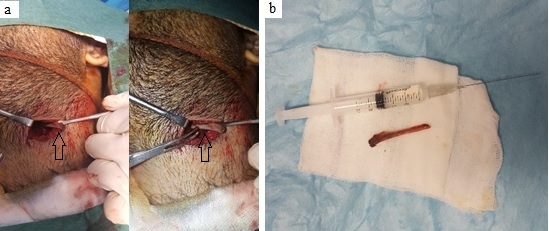
AbstractDespite its rarity, foreign body ingestion can result in severe complications such as pharyngoesophageal perforation, aortoesophageal fistula, carotid rupture, and deep neck infection. Patients with suspected foreign body ingestion should be carefully evaluated, and imaging techniques must be employed for diagnosis when required. In this article, we reported a 27-year-old man presented with deep neck infection and an ingested foreign body (chicken bone) was removed from his cervical abscess.IntroductionForeign body ingestion is one of the most frequent emergencies for which emergency departments request consultations from ear, nose, and throat physicians. Foreign bodies in the oropharyngeal region are frequently seen in childhood, although adult patients are also encountered. Fish bones are most commonly observed oropharyngeal foreign bodies in adults [1,2]. Complications such as pharyngoesophageal perforation, aortoesophageal fistula, carotid rupture, and deep neck infection, which are rare but may be life-threatening if not treated promptly, may occur in association with foreign body ingestion [3]. Patients presenting with a history of foreign body being ingested or trapped in the throat must therefore be treated with care. We report a case of a foreign body (chicken bone) passing from the pharyngeal region to the parapharyngeal area in a patient presenting with swallowing and breathing difficulty, and with cervical swelling. Case ReportA 27-year-old man with no known previous disease presented to our clinic with pain in the left side of the neck, increasing swelling, and worsening difficulty in swallowing and breathing persisting for approximately the preceding one week. The patient was hospitalized with a preliminary diagnosis of parapharyngeal abscess. Trismus was present at examination, with oral opening being measured at 1.5 cm. The oral cavity, teeth, tongue, and oropharynx were normal in appearance at endoscopic examination. At laryngoscopic examination, the laryngeal mucosa were healthy in appearance, and the bilateral vocal cords were mobile. However, the left pharyngeal wall exhibited marked medial displacement and was obstructing the airway. Painful swelling in the left submandibular region was observed at head and neck examination. Body temperature was high, and marked white cell elevation at complete blood count was also observed. Radiological imaging with computed tomography (CT) of the neck revealed irregular air densities and a hyperdense area suggestive of a foreign body inside the abscess pouch at the level of the hyoid bone in the left parapharyngeal region (Figure 1).
DiscussionForeign body ingestion is an emergency condition frequently encountered across the world. Children constitute 80% of cases, and cases peak between the ages of six months and three years. Adults represent a much smaller proportion of cases, and predisposing factors such as mental disability, immunosuppression, diabetes, and false teeth use may be present in some [4,5]. The most commonly involved foreign bodies in adults are fish bones, in approximately 70% of cases [6]. In the present case, however, although no specific information was obtained from the patient’s history, we identified a quite large chicken bone in the parapharyngeal region. CT is an effective tool in the diagnosis and location of deep neck abscesses and foreign bodies, and provides useful information from the spread of infection to the surgical approach [3]. When the patient in the present case, admitted with a preliminary diagnosis of parapharyngeal abscess, was evaluated using CT, we suspected that the foreign body had progressed to the parapharyngeal region. Pharyngeal foreign bodies are frequently encountered in the literature, but migration from the pharynx to the parapharyngeal region is rare. We think that contraction of the neck muscles may have assisted the displacement of the foreign body. Emergency intervention is essential in foreign bodies reaching an area contiguous to the carotid since these may damage the carotid wall and pose a severe risk of bleeding [2,7]. In the present case, the foreign body, which was in close proximity to the carotid, was approached with external exploration, and the body was carefully removed from the vicinity of the carotid. İn conclusion, due to the severe complications that may result from foreign body ingestion, such cases must undergo detailed head and neck examination, and intervention must be planned in the early period. Imaging techniques provide useful information, from diagnosis to the therapeutic approach. The possibility of a foreign body should be remembered in cervical and parapharyngeal masses. Informed ConsentFrom the patientReferences
Presented at3. Doğu Anadolu Acil Tıp Günleri, 04-06.10.2019, Erzurum |
|||||
| Keywords : Parafaringeal Yabancı Cisim , Parafarengeal Apse , Tavuk Kemiği , Derin boyun enfeksiyonu | |||||


|