|
UNILATERAL SECONDARY MIDDLE TURBINATE WITHOUT SINUSITIS: A RARE CLINICAL ENTITY
UNILATERAL SECONDARY MIDDLE TURBINATE WITHOUT SINUSITIS: A RARE CLINICAL ENTITY
Articles >
Rhinology
Submitted : 31.07.2023
Accepted : 04.10.2023
Published: 04.10.2023
|
|
Abstract
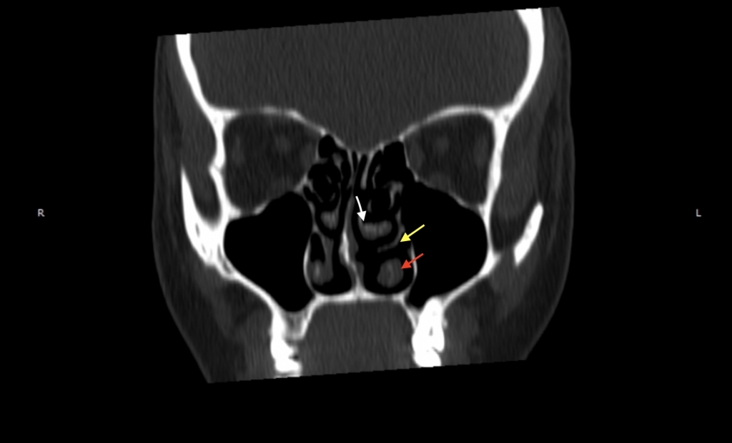
The anatomy and embryology of the nasal cavity and paranasal sinuses are quite complex. Nasal turbinates are important anatomical structures that extend from the lateral nasal wall to the nasal cavity. After the widespread use of endoscopes and computed tomography, many variations of these structures have been described. The secondary middle turbinate is a one of the rare variations of the nasal cavity. It is a soft tissue-covered bony prominence arising from the lateral nasal wall. It usually causes sinusitis by closing the osteomeatal complex. We present the case of a female patient diagnosed with this rare anatomical variation by means of computed tomography. On examination, a mass thought to be a nasal polyp was detected in the left nasal cavity. The computed tomography of the paranasal sinuses was done which showed a bony structure attached to the lateral wall of the middle meatus and covered by a soft tissue. It diagnosed as secondary middle turbinate and then the patient was treated with surgical intervention. Accurate identification of the anatomical variations of the turbinates will help to determine the correct diagnosis and treatment and to prevent possible complications during endoscopic procedures.
|
|
Keywords:
Nasal turbinate
, anatomical variation
, secondary middle turbinate
, otorhinolaryngology.
|
Özet
Burun boşluğu ve paranazal sinüslerin anatomisi ve embriyolojisi oldukça karmaşıktır. Burun konkaları, burun yan duvarından burun boşluğuna kadar uzanan önemli anatomik yapılardır. Endoskopların ve bilgisayarlı tomografinin yaygınlaşmasından sonra bu yapıların birçok varyasyonu tanımlanmıştır. İkincil orta konka, burun boşluğunun nadir varyasyonlarından biridir. Burun yan duvarından kaynaklanan yumuşak doku kaplı kemik çıkıntıdır. Genellikle osteomeatal kompleksi kapatarak sinüzite neden olur. Bu olgu sunumunda; bu nadir anatomik varyasyonun bilgisayarlı tomografi ile teşhis edildiği kadın hastayı sunuyoruz. Muayenede sol burun boşluğunda nazal polip olduğu düşünülen kitle tespit edildi. Paranazal sinüslerin bilgisayarlı tomografisinde orta mea yan duvarına yapışık ve yumuşak bir doku ile örtülü kemiksi bir yapı görüldü. Sekonder orta konka olarak teşhis edildi ve sonrasında hasta cerrahi müdahale ile tedavi edildi. Konkaların anatomik varyasyonlarının doğru tanımlanması, endoskopik işlemler sırasında doğru tanı ve tedavinin belirlenmesine ve olası komplikasyonların önlenmesine yardımcı olacaktır.
|
|
Anahtar kelimeler:
Nazal konka
, anatomik varyasyon
, ikincil orta konka
, kulak burun boğaz.
|
|
|
|