

Authors
|
|||||||||||
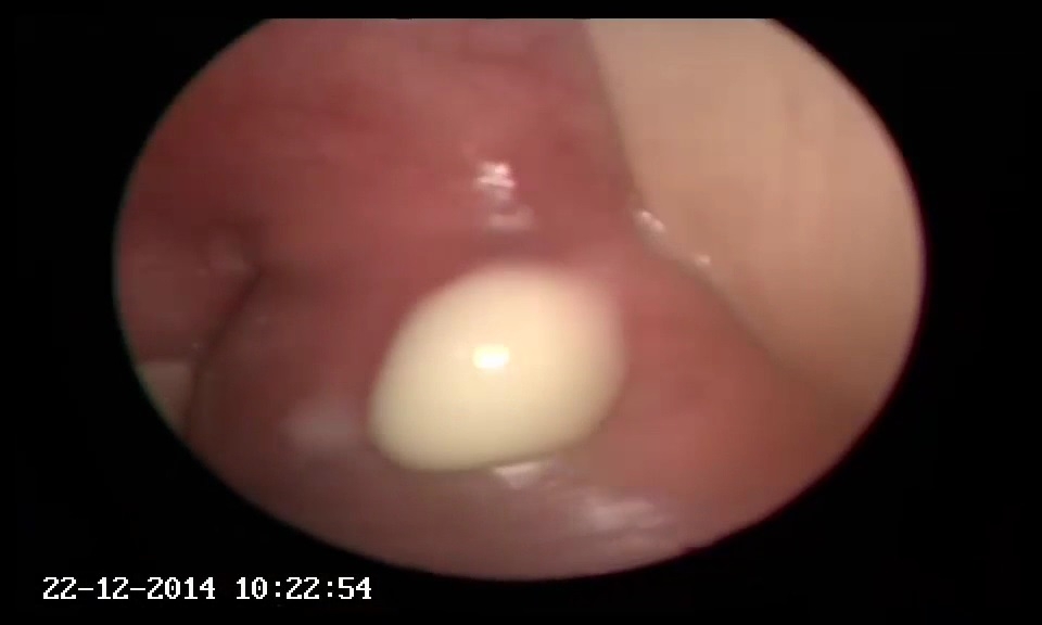
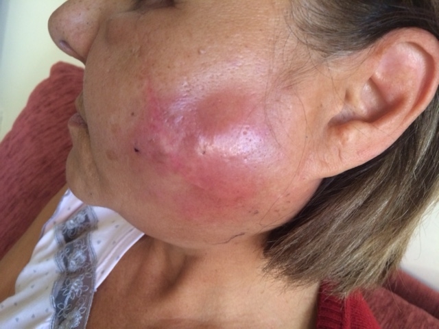
AbstractRecurrent acute parotitis without total resolution is called chronic parotitis. Acute parotitis is usually caused by viral or bacterial infection. There have been few cases reported with foreign body insertion to parotid gland causing chronic inflammation. Surgery and removal of the foreign body is the only treatment, care must be taken not to cause any facial nerve and Stensen's duct injuries. We present a rare case of chronic sialoadenitis as a consequence of multiple foreign bodies in a 63 year-old female which had most probably entered through the skin during a traffic accident 20 years ago.IntroductionChronic sialoadenitis is a disease of salivary glands with course of inflammation and pain episodes, generally caused by decreased salivary flow or stasis. It is mostly encountered in parotid gland [1]. Infective or obstructive sialoadenitis may present with pain and swelling of the affected salivary gland. Decreased salivary flow or stasis causes obstructive sialoadenitis. Stones obstructing salivary duct, strictures, tumors or rarely foreign bodies can cause stasis, which induces formation of obstructive sialoadenitis [2]. Sialolithiasis forms 66% of obstructive salivary diseases [3]. Foreign bodies in parotid gland causing obstructive sialoadenitis are extremely rare. Only a few cases have been reported with foreign bodies in the parotid gland [4]. Most of them enter the gland via Stensen’s duct. Penetration through the skin is very seldom. Here we report a case with multiple foreign bodies in parotid gland which had most probably entered through the skin during a traffic accident 20 years ago. Case ReportA 63 year old female patient presented to our outpatient clinic with swelling about 4x4cm in left cheek in parotid gland location. Patient had no chronic diseases and no history of smoking but had a traffic accident about 20 years ago which had injured left side of her face and had huge laceration involving parotid gland. Her physical examination showed that overlying skin was erythematous and tender. Oral cavity inspection revealed the presence of purulent secretion from the orifice of the parotid duct, after gentle pressure over the gland (Figure 1).
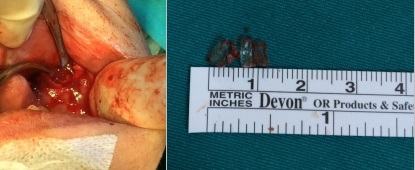
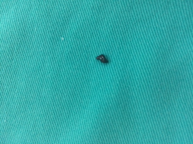
After taking consent of the patient, under general anesthesia, gland orifice was incised and using a probe, duct was dilated where multiple pieces of glass were removed gently (Figure 3).
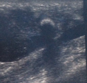
Patient had no complaints 3 months postoperatively and no foreign body or mass was seen in postoperative ultrasonography. DiscussionChronic inflammation of salivary glands is caused by chronic bacterial infection as a result of incomplete treatment or mistreatment of an acute disease. The disease frequently affects the parotid glands. Common pathogens are Staphylococcus aureus, Streptococcus viridans, St. pneumoniae, St. pyogenes and Escherichia coli. Streptococcus is the most commonly isolated agent in recurrent cases [5]. Viral and granulomatous and non-infectious parotitis can also be seen. Drugs such as diuretics, atropine and some antidepressants provoke inflammation by reducing salivary flow. Reduced salivary secretion and sialectasis are the most common findings of chronic inflammation [6]. Dehydration, malnutrition, chronic diseases, poor oral hygiene, and trauma increase the risk of parotitis [7]. Apart from stones, obstructive sialoadenitis may be due to calculi, mucinous lesions, tumors or anatomic variations or malformations of the duct [8], which cause stenosis of the duct. Foreign bodies are also a cause for obstructive sialoadenitis. Few cases have been reported with foreign bodies in the salivary glands, which include wood, grass, feather, pencil tip, toothbrush, fishbone, hair and finger nail [5]. Foreign bodies generally enter the parotid gland via Stensen’s duct, while penetration through the skin is seldomly seen [9]. The first case of foreign body in parotid gland was published in 1958 [10]. Penetrating accidents like gunshot and bullet in parotid gland have also been reported [11]. Foreign bodies left untreated or nondiagnosed cause complications that include prolonged or recurrent inflammation, abscess formation, cutaneous fistula, recurrent trismus [5]. Dilation of the salivary ducts as a consequence of sialolithiasis/foreign bodies can be easily diagnosed with ultrasonography. Ultrasonography is noninvasive and easy to apply for differantiation of glandular and extraglandular lesions, as well as cystic and solid lesions. Ultrasonography is also useful for biopsies and abscess drainage [12]. In our case ultrasonography showed the plastic body but was insufficent for the glass compartments. CT scan is an alternative option for diagnosing foreign bodies in salivary glands. Drainage of an abscess of the parotid gland should be performed very carefully in order to avoid any trauma to the facial nerve. Superficial parotidectomy may be an ultimate option for cure, but taking the complications of this method into account, less invasive and gland sparing methods are preferred [13]. Sialendoscopy is used in removal of lithiasis but there has been no report of it being used for foreign bodies [8]. We used external approach to parotid gland to reach the main foreign body and intraoral approach via Stensen’s duct to reach the glass components which could not be seen in ultrasonography but were palpated bimanually. This case is interesting not only because an obstructive foreign body in parotid gland is rare, but also it remind us how patient history is important and a foreign body may cause an obstructive sialoadenitis 20 years after penetrating the parotid gland. References
|
|||||||||||
| Keywords : Kronik sialoadenit , Yabancı cisim , Parotis bezi , Plastic , Cam | |||||||||||

|